41 lungs pictures with labels
Lung Anatomy, Function, and Diagrams - Healthline The lungs begin at the bottom of your trachea (windpipe). The trachea is a tube that carries the air in and out of your lungs. Each lung has a tube called a bronchus that connects to the trachea ... Chest anatomy illustrations - e-Anatomy - IMAIOS Anatomy of the chest and the lungs: anatomical illustrations. This e-Anatomy module presents an illustrated anatomy of the lungs, trachea, bronchi, pleural cavity and pulmonary vessels. This thoracic and pulmonary anatomy tool is especially designed for students of anatomy (medical and paramedical studies).
Label Lungs Diagram Printout - EnchantedLearning.com Human Anatomy. Read the definitions below, then label the lung anatomy diagram. bronchial tree - the system of airways within the lungs, which bring air from the trachea to the lung's tiny air sacs (alveoli). cardiac notch - the indentation in the left lung that provides room for the heart. diaphragm - a muscular membrane under the lungs.

Lungs pictures with labels
Lungs: Anatomy, Function, and Treatment - Verywell Health Lung tissue diseases like pulmonary fibrosis and sarcoidosis. There are 30,000 to 40,000 new cases of pulmonary fibrosis diagnosed in the U.S. each year, affecting 100,000 people in total. Sarcoidosis is considered a rare disease, affecting fewer than 200,000 in the U.S. Lungs (Human Anatomy): Picture, Function, Definition, Conditions Lung Tests. Chest X-ray: An X-ray is the most common first test for lung problems.It can identify air or fluid in the chest, fluid in the lung, pneumonia, masses, foreign bodies, and other ... 200+ Free Lunge & Lungs Images - Pixabay 275 28. man smoking cigarette. 51 10. green tea lung ching. 102 20. lungs branches tree. 91 17. health medicine anatomy. 89 21.
Lungs pictures with labels. 360 Lungs and Chest ideas | lunges, human lungs, lungs art Aug 24, 2015 - Explore Art In Biology's board "Lungs and Chest", followed by 174 people on Pinterest. See more ideas about lunges, human lungs, lungs art. Picture Illustration of Anatomical Structures - Lungs Picture of Lungs. The lungs are organs used for breathing located on either side of the chest. The lungs fill with air, oxygenate the blood, and dispose of carbon dioxide. Lungs are comprised of many different structures. The image on this page depicts the trachea, bronchi, and the several lobes of the left and right lungs. Labeled diagram of the lungs/respiratory system. - SERC View Original Image at Full Size. Labeled diagram of the lungs/respiratory system. Image 37789 is a 1125 by 1408 pixel PNG Uploaded: Jan10 14. Last Modified: 2014-01-10 12:15:34 Lungs Anatomy | Shapes and Surfaces of the Lungs 2. 3. A double-walled, fluid-filled sac called the pleura envelops each lung and aids in the ventilation process. 1. 2. The anterior, lateral, and posterior lung surfaces lie adjacent to the ribs and are thus often referred to as the costal surface.
Respiratory System Labelled Diagram Display Poster | Twinkl This model of the respiratory system labelled with the names of all the relevant parts is a great tool to teach children about the lungs in the human body, breathing, and the essential functions of the respiratory system. The handy labelled diagram highlights the main parts of the respiratory system, such as the trachea, bronchi, bronchioles, alveoli and the diaphragm. There's also a ... This Is What COPD Looks Like in the Lungs - Verywell Mind Stocktrek Images / Getty Images. Chronic bronchitis is a condition in which the airways, or your bronchi, become inflamed. This leads to mucus build-up in the airways that gets progressively worse. The symptoms include: 2. Coughing spells. Coughing up mucus or phlegm. Feelings of breathlessness 3. Wheezing. Chest pain. The lobes of the lungs Images, Stock Photos & Vectors - Shutterstock Find The lobes of the lungs stock images in HD and millions of other royalty-free stock photos, illustrations and vectors in the Shutterstock collection. Thousands of new, high-quality pictures added every day. Human Lung Alveoli Photos and Premium High Res Pictures - Getty Images lung x-ray of a heart failure - human lung alveoli stock pictures, royalty-free photos & images. alveolar wall, alveoli & respiratory membrane. normal human lung tissue, 250x at 35mm. shows the thin alveolar wall (simple squamous epithelium) and the extremely thin respiratory membrane beautifully adapted for gas exchange. also, shows red blood ...
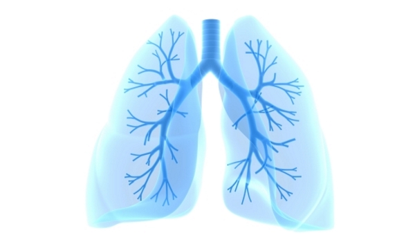
Labeled Diagram of the Human Lungs - Bodytomy Bronchi. The trachea or windpipe is the major structure that connects the nasal and oral cavities to the lungs. The trachea bifurcates into main branches called bronchi, which enter into the two lungs. The bronchi are made up of hyaline cartilage and smooth muscles. The left and right bronchi also differ in their dimensions, with the right one ... 100+ Best Lung Photos · 100% Free Download - Pexels Download and use 100+ Lung stock photos for free. Thousands of new images every day Completely Free to Use High-quality videos and images from Pexels. Explore. License. Upload. Upload Join. Lungs Doctor Breath Hospital Medical Health Air. Lung Pictures. Browse through our collection of LUNG pictures. All our photos are of high quality, so go ... Lung Cancer Pictures: CT Scan, X Ray, and More - Healthline CT scan. A computed tomography (CT) scan is often ordered if there is something abnormal on the chest X-ray. A CT scan takes a cross-sectional and a more detailed image of the lung. It can give ... Lungs Picture Image on MedicineNet.com The lungs are a pair of spongy, air-filled organs located on either side of the chest (thorax). The trachea (windpipe) conducts inhaled air into the lungs through its tubular branches, called bronchi.The bronchi then divide into smaller and smaller branches (bronchioles), finally becoming microscopic.The bronchioles eventually end in clusters of microscopic air sacs called alveoli.
BIO 202 lab- lungs
Lung Lobes: Definition, Anatomy, Functions, Picture Unlike the right lung, there are only two lobes in the left lung: the superior (upper) and inferior (lower) lung lobes [3]. Why does the right lung have three lobes and the left lung has two. The left lung is a little smaller than the right lung because it has to make space for the heart (the cardiac notch) in the left side of the thoracic cavity.

Dentistry and Medicine: Thorax,Lungs,Heart Anatomy and Physiology Diagrams Free Download
774 Label the lungs Vector Images, Label the lungs Illustrations ... 774 Royalty-free Vector Images & Drawings of Label the lungs. artskvortsova Lungs symbol. Breathing. Lunge exercise. Lung cancer. artskvortsova Lungs symbol. Breathing. Lunge exercise.

Sardonyx Healing Properties
Lung Anatomy Printable | Lung anatomy, Human body ... - Pinterest Lung Anatomy Printable. Description This easy and visual lung model is an easy way to teach kids lung anatomy and how lungs work to keep us breathing. Attach it to a straw with two plastic sandwich bags and have kids inflate in and out to see how lungs work. It makes a fun science anatomy craft or activity for kids homeschool or in the classroom!

Lungs on Behance
Human Lung Anatomy Photos and Premium High Res Pictures - Getty Images Browse 8,064 human lung anatomy stock photos and images available, or start a new search to explore more stock photos and images. lungs anatomy. human internal organ. - human lung anatomy stock illustrations. male lung, illustration - human lung anatomy stock illustrations.

Lungs Medical Exhibit
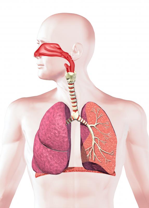
Lung Anatomy Diagram, Respiratory System Function Diagram of the respiratory system. Air enters the body via the nose (preferably) or the mouth. The air enters the main windpipe, called the trachea, and continues en route to each lung via either the right or left bronchus (plural=bronchi). The lungs are separated into sections called lobes, two on the left and three on the right.

Nursing Student Study Materials: picture of lungs
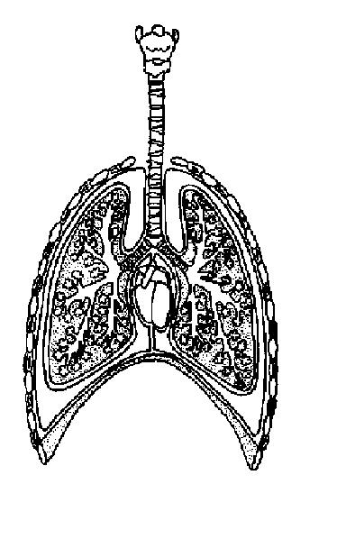
Lung Diagram - Pictures, Photos & Images of Biology - Science for Kids Find free pictures, photos, diagrams, images and information related to a wide range of different biology topics right here at Science Kids. Photo name: Lung Diagram Picture category: Biology Image size: 61 KB Dimensions: 785 x 600 Photo description: A black and white diagram of the human lungs.The image shows a brief outline of the human chest, the trachea or windpipe as well as the bronchial ...
Respiratory System Worksheet - WikiEducator
PDF ANATOMY OF LUNGS - University of Kentucky Lungs are a pair of respiratory organs situated in a thoracic cavity. Right and left lung are separated by the mediastinum. Texture-- Spongy Color - Young - brown Adults -- mottled black due to deposition of carbon particles Weight-Right lung - 600 gms Left lung - 550 gms.
What is Rheumatoid Arthritis of the Lung? (with pictures)
Lungs: Definition, Location, Anatomy, Function, Diagram, Diseases Where are the Lungs Located. The lungs are located a little toward the posterior part of the human body, just below the collarbone, extending down to the diaphragm, the muscular partition that separates the chest and abdominal cavities.The left and right lungs are situated on the two sides of the body with the heart, another vital organ in the thoracic cavity, located a little in front of, and ...

Lungs on Behance
900+ Lungs Clip Art | Royalty Free - GoGraph Abstract Futuristic Human Lungs Wireframe Blue Digital Point Connecting Concept Analysis And Diagnosis Of Pulmonary Diseases, Respiratory Disease, Lung Health, Medical Care For Patients. Pulmonologist Listens Body Lungs. Human Lung Anatomy. The Lungs Of Man Vintage Engraving.

The lungs - Stock Image - F002/0666 - Science Photo Library
Human Lung Stock Photos, Pictures & Royalty-Free Images - iStock Browse 61,510 human lung stock photos and images available, or search for human lung anatomy or human lung illustration to find more great stock photos and pictures. 3D illustration of Lungs, medical concept. Human lungs anatomy form lines and triangles, point connecting network on blue background.

Lung Facts, Anatomy, and Overview in Pictures
Diagram Of The Respiratory System With Labels Stock Photos, Pictures ... lung. The lungs are the primary organs of respiration in humans and many other animals including a few fish and some snails. In mammals and most other vertebrates, two lungs are located near the backbone on either side of the heart. Vector graphic. diagram of the respiratory system with labels stock illustrations

Lungs - Colored
200+ Free Lunge & Lungs Images - Pixabay 275 28. man smoking cigarette. 51 10. green tea lung ching. 102 20. lungs branches tree. 91 17. health medicine anatomy. 89 21.

ClearLungs® Extra Strength 120 Veg Capsules - Item 146
Lungs (Human Anatomy): Picture, Function, Definition, Conditions Lung Tests. Chest X-ray: An X-ray is the most common first test for lung problems.It can identify air or fluid in the chest, fluid in the lung, pneumonia, masses, foreign bodies, and other ...
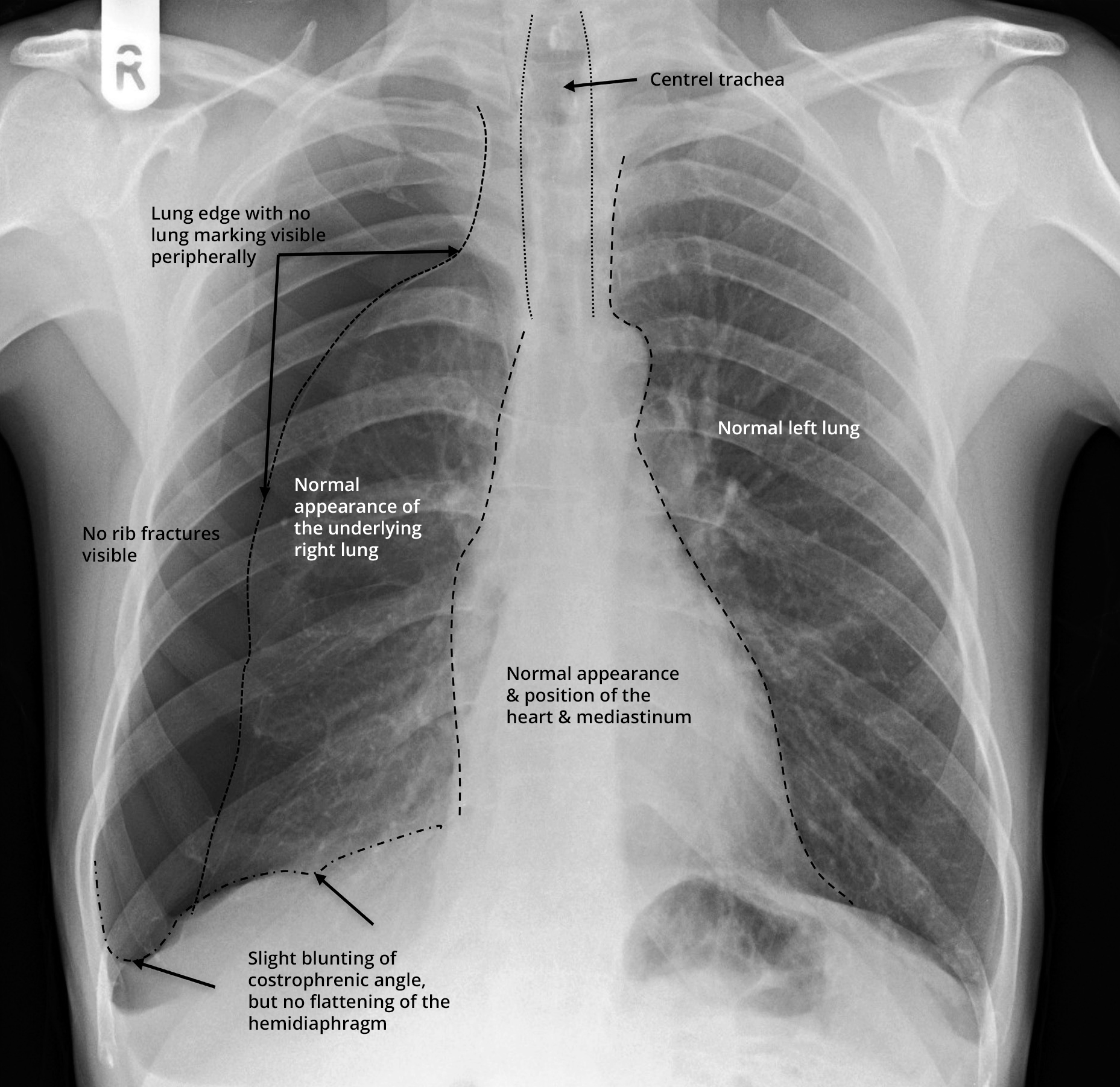
The Unofficial Guide to Radiology
Lungs: Anatomy, Function, and Treatment - Verywell Health Lung tissue diseases like pulmonary fibrosis and sarcoidosis. There are 30,000 to 40,000 new cases of pulmonary fibrosis diagnosed in the U.S. each year, affecting 100,000 people in total. Sarcoidosis is considered a rare disease, affecting fewer than 200,000 in the U.S.

Federal judge nixes graphic anti-smoking ads on cigarette packages - NY Daily News
The role of Alveoli in the respiratory system

The lungs - Stock Image - F002/0334 - Science Photo Library
Post a Comment for "41 lungs pictures with labels"