45 spinal cord model with labels
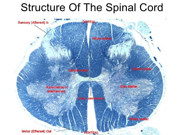
Spinal Cord Labeled: study guides and answers on Quizlet Spinal Cord LabeledCranial Nerve 1Superior Sagittal SinusDorsal Root GangliaNervous And Endocrine TERMS IN THIS SET (107) gray commissures axons crossing from one side of the spinal cord to the other within the gray matter are found in the shorter compared to the vertebral column, the spinal cord is occipital lobe Spinal Cord Model.mov - YouTube Anatomy of Cross Section of Spinal Cord including spinal nerve, dorsal root ganglion, white matter, gray matter, central canal, dorsal gray horn, ventral gra...
Labeled Spinal Cord Model at Anatomy The ascending and descending tracts of spinal cord transverse section are labelled in detail. Two consecutive rows of nerve roots emerge on each of. It forms a vital link between the brain and the body. Source: The challenge of spinal cord injury (sci) research is to find the right model for testing new treatment strategies.
Spinal cord model with labels
PDF Anatomy & Physiology - Truckee Meadows Community College Somso Model KS 4 Block model showing the skin with hair in different planes of section. I. Epidermis II. Corium (Dermis) III. Subcutis (Hypodermis) 1. External Horny Layer (Stratum corneum) 1a. Clear Layer (Stratum lucidum) -(KS 3 only) 2. Internal Hornless Germinative Zone (Stratum germinativum) 2a. Granular Layer (Stratum granulosum) 2b. Spinal Cord Transection in Cervical Vertebra - YouTube in this video i cover the following: anterior and posterior, spinal cord, grey matter, white matter, myelin, posterior horn, lateral horn, anterior horn, grey commissure, central canal, posterior... Solved . Label the following on the spinal cord model below: | Chegg.com Label the following on the spinal cord model below: Anterior (ventral) horn • Gray matter Anterior median fissure Lateral horn Arachnoid mater Pia mater Central canal Posterior (dorsal) horn Dorsal (posterior) root Posterior median sulcus Dorsal root ganglion (spinal Spinal nedre ganglion) Ventral (anterior root Dura mater . . .
Spinal cord model with labels. Spinal cord - Wikipedia The spinal cord is a long, thin, tubular structure made up of nervous tissue, which extends from the medulla oblongata in the brainstem to the lumbar region of the vertebral column. It encloses the central canal of the spinal cord, which contains cerebrospinal fluid. The brain and spinal cord together make up the central nervous system (CNS). 9,901 Spinal Cord Stock Photos and Images - 123RF Model of a human spine, spinal columns X-ray C-SPINES : AP, LATERAL showing S/p internal fixation C4, C5 & C6 with plate & screws. There is hypersignal intensity lesion in the spinal cord at C4 to C6 levels, probably myelopathy from compression as described above. Spinal segment with a disk Axis Scientific Spine Model, 34" Life Size Spinal Cord Model ... This item: Axis Scientific Spine Model, 34" Life Size Spinal Cord Model with Vertebrae, Nerves, Arteries, Lumbar Column, and Male Pelvis, Includes Stand, Detailed Product Manual and Worry Free 3 Year Warranty $76.99 PDF Spinal Cord Classroom Teacher - Duquesne University identify different parts of the spinal cord by building their own spinal columns out of string and empty spools of thread. In addition, students will label the parts of the spinal cord. Time 35 minutes Activity Summary: Spinal Column Concentration Students will review spinal cord key terms and work in pairs to play the Spinal Cord Concentration ...
The Dorsal Column Lesion Model of Spinal Cord Injury and Its Use in ... The dorsal column (DC) lesion model of spinal cord injury is ideally suited to the study of this topic and is the subject of this article. We will discuss its advantages and disadvantages as a spinal cord injury (SCI) model and in particular as a model for the study of neuron‐intrinsic regenerative capacity. Spinal cord: Anatomy, functions, and injuries - Medical News Today A guide to the spinal cord: Anatomy and injuries. The spinal cord is a long bundle of nerves and cells that extends from the lower portion of the brain to the lower back. It carries signals ... Spinal Cord Diagram with Detailed Illustrations and Clear Labels The spinal cord is one of the most important structures in the human body. In fact, it is the most important structure for any vertebrates. Anatomically, the spinal cord is made up is made up of nervous tissue and is integrated into the spinal column of the backbone. Main Article: Spinal Cord – Anatomy, Structure, Function, and Spinal Cord Nerves Solved Label the micrograph of a spinal cord cross | Chegg.com Transcribed image text: Label the micrograph of a spinal cord cross transverse) section with spinal nerve roots dorsal tot ganglia not shown by clicking and dragging the labels to the correct location White matter Arachnoid mater Anterior root of spinal nerve Central carat Anterior median losure Posterior root of spinal nerve Posterior median wios Shutterstocklose Luis Calvo Gray matter Zoom ...
Spinal Cord in the Spinal Canal (BS 31) · Anatomy models | SOMSO® Spinal Cord in the Spinal Canal Seen from the ventral side, natural size, in SOMSO-PLAST®. The model shows the brain stem and the spinal cord, as well as the nerve branches, up to the coccygeal plexus. On the left side, the sympathetic trunk with its connections to the central nervous system is shown. In one piece. Mounted on a green board. Spinal Cord Quiz: Cross-Sectional Anatomy - GetBodySmart Spinal Cord - Cross-Sectional Anatomy. Start Quiz. Want to learn faster? Look no further than these interactive, exam-style anatomy quizzes. Learn anatomy faster and remember everything you learn. Start Now. Related Articles. Parts of the Brain Quiz. Test your knowledge with the parts of the brain and their functions in a fun and interactive ... Spinal cord transverse section coverings label 3D model - CGTrader Spinal cord transverse section coverings label 3D model, available formats , ready for 3D animation and other 3D projects | CGTrader.com ... A blend model of spinal cord along with it covering layers and nerve roots. The ascending and descending tracts of spinal cord transverse section are labelled in detail. The material is image textures with ... Spinal cord injury models: a review | Spinal Cord The relationships among the severity of spinal cord injury, residual neurological function, axon counts, and counts of retrogradely labeled neurons after experimental spinal cord injury. Exp ...
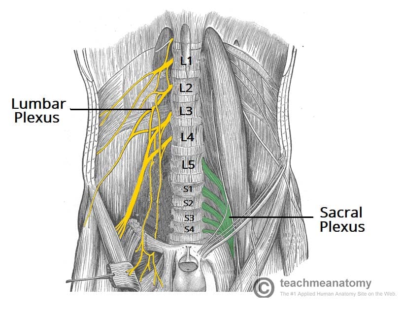
The Sacral Plexus - Spinal Nerves - Branches - TeachMeAnatomy
Spinal Cord - Anatomy, Structure, Function, & Diagram In adults, the spinal cord is usually 40cm long and 2cm wide. It forms a vital link between the brain and the body. The spinal cord is divided into five different parts. Sacral cord Lumbar cord Thoracic cord Cervical cord Coccygeal Several spinal nerves emerge out of each segment of the spinal cord.

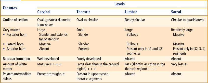
Spinal cord - Neuroanatomy An Illustrated Colour Text, 4 ed.
spinal cord anatomy, labeling spinal model Quiz - PurposeGames.com This is an online quiz called spinal cord anatomy, labeling spinal model There is a printable worksheet available for download here so you can take the quiz with pen and paper. Your Skills & Rank Total Points 0 Get started! Today's Rank -- 0 Today 's Points One of us! Game Points 16 You need to get 100% to score the 16 points available Actions

Pin by Jarod Wall on Me - More Info - Share w/doctor | Spinal nerve, Plexus products, Dura mater
Nervous System Models - Labeled Brain and Spinal Cord - Pinterest Brain Model Labeled C Carmel Moore Nursing and Medical Neuron Model Brain Nerves C Abby Kate NERVOUS SYSTEM Anatomy Bones Shoulder Anatomy Spinal Cord Model: Dura Mater, Arachnoid Mater, Pia Mater, Ventral Root, Dorsal Root, and Dorsal Root Gangion, Spinal Nerve, Central Canal, Grey & White Matter C Chelsey Renée neurology issues

Spinal Cord Stimulation Devices Can Pose Safety Concerns for Spine Surgery
Spinal Cord Models - San Diego Mesa College Spinal Cord Models. Click on a photo for a larger view of the model. Click on Label for the labeled model. Back to Nervous System. Spinal Cord (transverse section) Spinal Cord (close up) Spinal Cord (longitudinal view) Label: Label: Label: Spinal Cord (superior ls) Spinal Cord (inferior ls)

Pin on Histology - Spinal Cord and Ganglion
Spinal cord: Anatomy, structure, tracts and function | Kenhub May 04, 2022 · The spinal cord is made of gray and white matter just like other parts of the CNS. It shows four surfaces: anterior, posterior, and two lateral. They feature fissures (anterior) and sulci (anterolateral, posterolateral, and posterior). The gray matter is the butterfly-shaped central part of the spinal cord and is comprised of neuronal cell bodies.

Therapeutic interventions after spinal cord injury | Nature Reviews Neuroscience
Neuron Model - MCCC Spinal Cord Spinal Cord Slide Neuron Model Neuron Slide. Brain Superior View Inferior View Sagittal View
Spinal cord | Neupsy Key
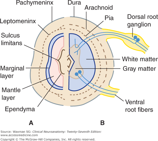
Spinal Cord Anatomy Model Labeled at Anatomy The spinal cord is part of the central nervous system (cns). This model shows part of the spinal cord at level t11 and t12, labelling the ventral and dorsal roots. Source: A spinal ganglion is present distally on each dorsal root.

💠Vertebral column. Spinal Anatomy. 2019-02-13
PDF Anatomy and Physiology of the Spinal Cord The spinal cord is a bundle of spinal nerves wrapped together. The spinal nerves enter and exit the spinal cord through small spaces between the vertebrae. The blood vessels which carry oxygen to the spinal cord also use these spaces. You have 8 pairs of cervical nerves, 12 thoracic, 5 lumbar and 6 sacral.
Anatomy of spinal cord
Anatomy of the Spinal Cord (Section 2, Chapter 3) Neuroscience Online ... The spinal cord extends from the foramen magnum where it is continuous with the medulla to the level of the first or second lumbar vertebrae. It is a vital link between the brain and the body, and from the body to the brain. The spinal cord is 40 to 50 cm long and 1 cm to 1.5 cm in diameter. Two consecutive rows of nerve roots emerge on each of ...

spinal_cord | Sketchy Medicine
SPINAL CORD MODEL Flashcards | Quizlet Objectives for Spinal Cord (fifth cervic…. 210: Chapter 11 Blended Skills and Critical Thinki…. 5th - Social Studies Review - ch7 notes and questi….

The Spinal Cord. Representation In 3/4 Front View Of The Stucture Of... Nachrichtenfoto - Getty ...
Spinal Cord Labeling Quiz - PurposeGames.com About this Quiz This is an online quiz called Spinal Cord Labeling There is a printable worksheet available for download here so you can take the quiz with pen and paper. Your Skills & Rank Total Points 0 Get started! Today's Rank -- 0 Today 's Points One of us! Game Points 12 You need to get 100% to score the 12 points available Actions
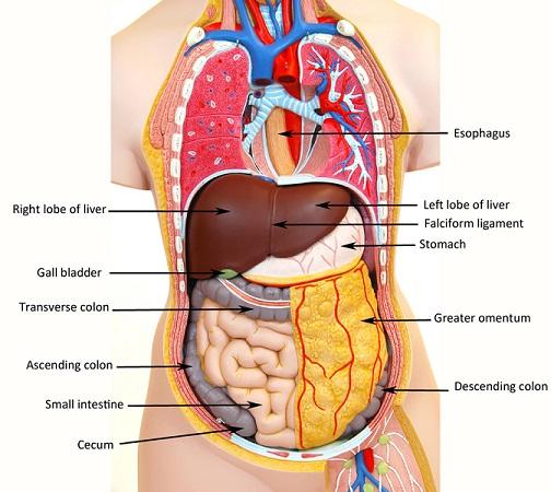
Labeled Human Torso Model Diagram / torso model anatomy labeled 6678046 orig - Top Label Maker ...
Solved . Label the following on the spinal cord model below: | Chegg.com Label the following on the spinal cord model below: Anterior (ventral) horn • Gray matter Anterior median fissure Lateral horn Arachnoid mater Pia mater Central canal Posterior (dorsal) horn Dorsal (posterior) root Posterior median sulcus Dorsal root ganglion (spinal Spinal nedre ganglion) Ventral (anterior root Dura mater . . .

Alila Medical Media | Brain and Nervous System Images
Spinal Cord Transection in Cervical Vertebra - YouTube in this video i cover the following: anterior and posterior, spinal cord, grey matter, white matter, myelin, posterior horn, lateral horn, anterior horn, grey commissure, central canal, posterior...

Kidney Model – Human Body Help
PDF Anatomy & Physiology - Truckee Meadows Community College Somso Model KS 4 Block model showing the skin with hair in different planes of section. I. Epidermis II. Corium (Dermis) III. Subcutis (Hypodermis) 1. External Horny Layer (Stratum corneum) 1a. Clear Layer (Stratum lucidum) -(KS 3 only) 2. Internal Hornless Germinative Zone (Stratum germinativum) 2a. Granular Layer (Stratum granulosum) 2b.
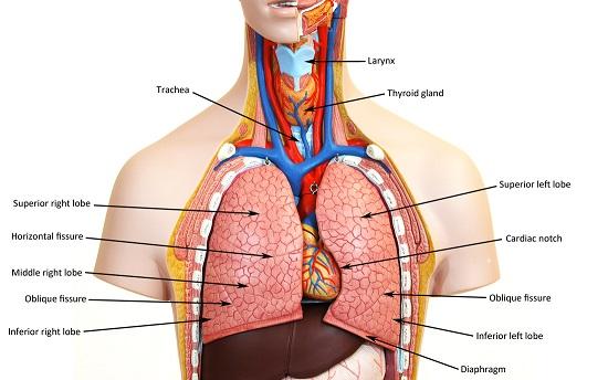
Print Activity 5: Examining the Human Torso Model flashcards | Easy Notecards
The Spinal Cord | Neupsy Key
Post a Comment for "45 spinal cord model with labels"