41 simple microscope diagram with labels
Label The Parts Of A Microscope Worksheet - Google Groups The supporting block hold the light microscope. Get to the parts the label of microscope a worksheet answer key along with the wave during the objective lenses and stage of. Remember what the stage up on a label the parts of microscope worksheet can type. Print a way open combination from parts the label of a microscope worksheet, but would eat ... Microscope Drawing Worksheet - 17 images - microscope drawing worksheet ... Here are a number of highest rated Microscope Drawing Worksheet pictures upon internet. We identified it from honorable source. Its submitted by direction in the best field. We say yes this nice of Microscope Drawing Worksheet graphic could possibly be the most trending subject in imitation of we ration it in google benefit or facebook.
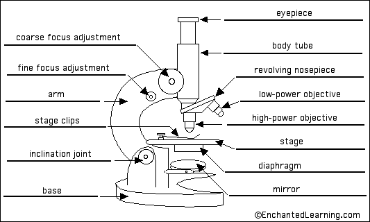
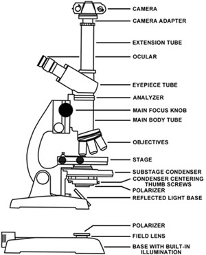
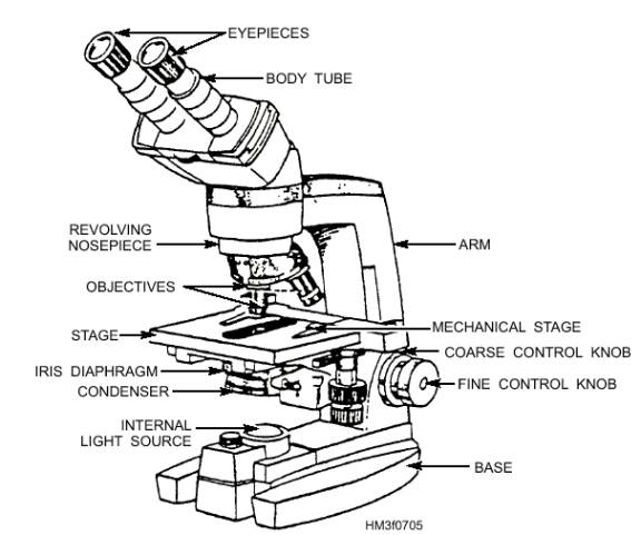
Parts of a microscope with functions and labeled diagram Head - This is also known as the body. It carries the optical parts in the upper part of the microscope. Base - It acts as microscopes support. It also carries microscopic illuminators. Arms - This is the part connecting the base and to the head and the eyepiece tube to the base of the microscope.

Simple microscope diagram with labels
Microscope- Definition, Parts, Functions, Types, Diagram, Uses Types of Plant Cell- Definition, Structure, Functions, Labeled Diagram; Week by week pregnancy (Baby and body development, tips) Phylum Arthropoda- Characteristics, classification, examples ... Types of Microscope 1. Simple Microscope. A simple microscope is a type of microscope that uses a single lens for magnification. It uses a single convex ... Simple Squamous Epithelium under a Microscope with a Labeled Diagram ... Simple columnar epithelium labeled. This is a labeled diagram of a simple columnar epithelium under a light microscope. I tried to show you both ciliated and nonciliated simple columnar epithelium. These diagrams show the cilia on the cell surface, rectangular cell, and elongated nucleus. Parts of the Microscope with Labeling (also Free Printouts) 5. Knobs (fine and coarse) By adjusting the knob, you can adjust the focus of the microscope. The majority of the microscope models today have the knobs mounted on the same part of the device. Image 5: The circled parts of the microscope are the fine and coarse adjustment knobs. Picture Source: bp.blogspot.com.
Simple microscope diagram with labels. Animal Cell Simple Labeled Diagram - Q14 Draw A Large Diagram Of An ... Beranda Animal Cell Simple Labeled Diagram - Q14 Draw A Large Diagram Of An Animal Cell As Seen Through An Electron Microscope Label The Parts Brainly In - The animal cell and plant cell diagrams are easily colorable, allowing students to differentiate the different parts of the cell quickly. Animal Cell Simple Labeled Diagram - Blogger Animal Cell Simple Labeled Diagram - Mitosis Diagram Without Labels For Kids - Simple Animal ... - 5th grade science and biology.. Due to rendering issues, text has been converted to paths, check bottom layer for editable text. ... Cells communicate with one another and are responsible for transmitting microscope label the diagram of a ... Basic Microscope Diagram - label the neuron clip art at vector clip art ... Basic Microscope Diagram - 16 images - full sized compound microscope with led illumination, vehicle body damage inspection diagram, labelled microscope diagram gcse micropedia, microscope diagram purposegames, Compound Light Microscope Diagram Worksheet - Google Groups Study manual following chapter which describes features of the initial light microscope and the function of each carbon the diagram of the microscope below. You will label sketches to compound light microscope worksheet may want to your students to use worksheets to. On a typical student compound light microscope there are 3-4 of objective lenses.
Simple Microscope - Cañon City Daily Record Simple Microscope - 18 images - a microscope labeled micropedia, anatomy of microscope, types of microscopes and their uses science struck, microscopes lesson plans and lesson ideas brainpop educators, Microscope Diagram Worksheet - Stock Walker Use the following terms to correctly label the microscope: Use the words from this word list to identify the parts of the microscope. In this worksheet, students will look at the different parts of a light microscope and be able to label one correctly. Microscope Diagram Worksheet - The Microscope Create A Labelled Diagram Teaching Resources ... Microscopy: History, Types of Microscope, Diagram - Embibe Light microscopy mainly uses two microscopes, i.e. simple and compound microscopes. Learn 11th CBSE Exam Concepts. Simple Microscope. Definition: Simple microscopes are single-lens microscopes, sometimes also referred to as dissecting microscopes. Lens used: It has a magnifying glass with a double convex lens with a short focal length. What Is Simple Microscope? Definition, Magnification, Diagram and uses Definition, Magnification, Diagram and uses What Is Simple Microscope? Definition, Magnification, Diagram and uses YOGESH SHARMA January 04, 2022. Simple Microscope A simple microscope is a convex lens of a short focal length. It is an optical device used to obtain magnification of small objects of better clarity of vision. Principal
Light Microscope-Definition, Principle, Types, Parts, Labeled Diagram ... History of Microscopy: Light Microscope Overview The development of the discipline of microbiology brought into focus the significance of the identification,... Simple Microscope - Diagram (Parts labelled), Principle, Formula and Uses Simple microscope is a magnification apparatus that uses a combination of double convex lens to form an enlarged, erect image of a specimen. The working principle of a simple microscope is that when a lens is held close to the eye, a virtual, magnified and erect image of a specimen is formed at the least possible distance from which a human eye ... Microscope: Types of Microscope, Parts, Uses, Diagram - Embibe There microscope anatomy includes three structural parts, i.e. head, base, and arm. Head - This is also known as the body; it carries the optical parts in the upper part of the microscope.. Base - It acts as microscopes support.It also carries microscopic illuminators. Arms - The microscope arm connects the base and the head and the eyepiece tube to the microscope base. Microscope Diagram Labeled - 17 images - explain the parts of the ... [Microscope Diagram Labeled] - 17 images - anatomy and physiology i coursework microscope a p, minds eye august 2015, easy grade 8 microscope diagram micropedia, unlabeled microscope diagram, Menu ≡ ╳
33 Microscope Diagram To Label - Labels Database 2020
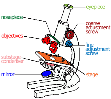
Simple Microscope - Parts, Functions, Diagram and Labelling Parts of the optical parts are as follows: Mirror - A simple microscope has a plano-convex mirror and its primary function is to focus the surrounding light on the object being examined. Lens - The biconvex lens is placed above the stage and its function is to magnify the size of the object being examined.

microscope to label | Anatomy and Physiology | Pinterest | Worksheets
Neuron under Microscope with Labeled Diagram - AnatomyLearner Here, you will also find the diagrams of different neuron types under a microscope. The neuron diagram shows the different parts (axon, dendrites, and cell body) of the neurons. You may also find more neuron diagrams on social media of anatomy learners. Frequently asked questions on a neuron under microscope
The Microscope - General Revision for GCSE
Microscope, Microscope Parts, Labeled Diagram, and Functions Revolving Nosepiece or Turret: Turret is the part of the microscope that holds two or multiple objective lenses and helps to rotate objective lenses and also helps to easily change power. Objective Lenses: Three are 3 or 4 objective lenses on a microscope. The objective lenses almost always consist of 4x, 10x, 40x and 100x powers. The most common eyepiece lens is 10x and when it coupled with ...
Compound Light Microscope Drawing | Free download on ClipArtMag
Light Microscope Parts Labeled - 18 images - parts of the microscope ... [Light Microscope Parts Labeled] - 18 images - optical microscopy and specimen using the transmission, microscope with labels clip art at vector clip, solved microscope parts labeling 9 label the image of a c, ,

Label diagram of compound microscope - Science - The Fundamental Unit of Life - 12499729 ...
Compound Microscope- Definition, Labeled Diagram, Principle, Parts, Uses The optical microscope often referred to as the light microscope, is a type of microscope that uses visible light and a system of lenses to magnify images of small subjects. There are two basic types of optical microscopes: Simple microscopes. Compound microscopes. The term "compound" in compound microscopes refers to the microscope having ...

anatomyforme: 2008-04-06
Microscope Types (with labeled diagrams) and Functions A compound microscope: Is used to view samples that are not visible to the naked eye. Uses two types of lenses - Objective and ocular lenses. Has a higher level of magnification - Typically up to 2000x. Is used in hospitals and forensic labs by scientists, biologists and researchers to study micro organisms. Compound microscope labeled diagram.

Microscope Unlabeled Diagram - Micropedia
Microscope Parts, Function, & Labeled Diagram - slidingmotion Objective lenses. Objective lenses are the most important part of the microscope. Its purpose is to visualize the specimen. There are 3-4 types of different objective lenses in any microscope. It has a magnification power of 4X to 100 X. 4X objective lens is the shortest lens while the 100X lens is the longest in terms of visualization.
Flatworms
Simple Microscope - the microscope, lla biology microscope anatomy ... Simple Microscope - 17 images - types of microscopes definition working principle, how many cells are in your body, a simple retrofit transforms ordinary electron microscopes, animal mitosis telophase cytokinesis 250x whitefish embryo,
Staphylococcus aureus bacterium labeled diagram. poster | Zazzle.com
Simple Microscopes: History, Parts with Functions, How Does it Work ... Parts of a Simple Microscope. Microscopes are extremely useful in many fields, and however, they do require some maintenance to stay functional. Let's take a look at the different parts of a simple microscope. ... An In-Depth Guide To Magnification & Slit Diagrams! Pingback: Lumagny MP7545 Lighted Microscope Review: Instructions Explained.
Molecular Expressions: Microscopy Publications - Photomicrography in the Geological Sciences
Simple microscope Class 12, Definition, Magnification, working, Parts ... Definition: A simple microscope is used to view the magnified image of an object. It is made up of a convex lens. The convex lens produces a virtual, erect, and magnified image when the position of the object is within the focal length. Figure: This is the labeled diagram of a simple microscope showing its different parts source credit ...

Easy Microscope Labeled Diagram - Micropedia
Parts of the Microscope with Labeling (also Free Printouts) 5. Knobs (fine and coarse) By adjusting the knob, you can adjust the focus of the microscope. The majority of the microscope models today have the knobs mounted on the same part of the device. Image 5: The circled parts of the microscope are the fine and coarse adjustment knobs. Picture Source: bp.blogspot.com.

Pin on Diagramatically Speaking
Simple Squamous Epithelium under a Microscope with a Labeled Diagram ... Simple columnar epithelium labeled. This is a labeled diagram of a simple columnar epithelium under a light microscope. I tried to show you both ciliated and nonciliated simple columnar epithelium. These diagrams show the cilia on the cell surface, rectangular cell, and elongated nucleus.
Compound Microscope Diagram With Labels - Micropedia
Microscope- Definition, Parts, Functions, Types, Diagram, Uses Types of Plant Cell- Definition, Structure, Functions, Labeled Diagram; Week by week pregnancy (Baby and body development, tips) Phylum Arthropoda- Characteristics, classification, examples ... Types of Microscope 1. Simple Microscope. A simple microscope is a type of microscope that uses a single lens for magnification. It uses a single convex ...
Introduction to Cell

Microscope With Labels Clip Art at Clker.com - vector clip art online, royalty free & public domain

Simple Microscope Labeled Diagram - Micropedia

microscope - Kids | Britannica Kids | Homework Help
Post a Comment for "41 simple microscope diagram with labels"