45 diagram of a human cell with labels
Cell: Structure and Functions (With Diagram) - Biology Discussion Eukaryotic Cells: 1. Eukaryotes are sophisticated cells with a well defined nucleus and cell organelles. 2. The cells are comparatively larger in size (10-100 μm). 3. Unicellular to multicellular in nature and evolved ~1 billion years ago. 4. The cell membrane is semipermeable and flexible. 5. These cells reproduce both asexually and sexually. human eye diagram with labels Labeled Volvox Diagram - Made By Creative Label labels-creative.com. volvox diagram labeled microscope cell under labels pandorina gonium label vegetative single creative chlorophyta. 35 Label The Structure Of The Eye - Labels Database 2020 otrasteel.blogspot.com. label eye labeling quiz structure associated lacrimal apparatus structures labels ...
PDF Human Cell Diagram, Parts, Pictures, Structure and Functions Diagram of the human cell illustrating the different parts of the cell. Cell Membrane The cell membraneis the outer coating of the cell and contains the cytoplasm, substances within it and the organelle. It is a double-layered membrane composed of proteins and lipids.

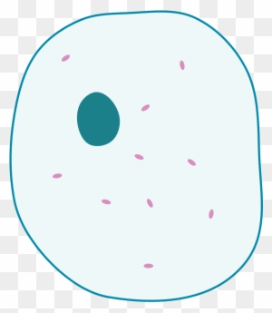
Diagram of a human cell with labels
A multi-omic single-cell landscape of human gynecologic ... Dec 02, 2021 · Deconvolution of regulatory mechanisms that drive transcriptional programs in cancer cells is key to understanding tumor biology. Herein, we present matched transcriptome (scRNA-seq) and chromatin accessibility (scATAC-seq) profiles at single-cell resolution from human ovarian and endometrial tumors processed immediately following surgical resection. Human Cells Printables and Diagrams - The Successful Homeschool These cells include: leukocytes, haematids, thrombocytes, ovum, sperm, sarcomeres, enterocytes, neurons, osteocytes, hepatocytes. They will learn the parts of a cell thanks to a labeled diagram. They will get to see what blood looks like under a microscope without needing to own a microscope. They get to color a cell and then label the parts. human cell diagram to label Histology basal cell deferens vas label anatomy ansci wisc edu male. Skin histology tissue connective types basic adipose components structure tissues layers system integumentary stain showing dermis dense epidermis irregular embryology. Cell human structure cells cellule nucleus parts anatomy nutshell dna noyau stuff human cell diagram to label
Diagram of a human cell with labels. 03 Label the Cell Diagram | Quizlet Start studying 03 Label the Cell. Learn vocabulary, terms, and more with flashcards, games, and other study tools. Labeled Diagram of the Human Kidney - Bodytomy Labeled Diagram of the Human Kidney The human kidneys house millions of tiny filtration units called nephrons, which enable our body to retain the vital nutrients, and excrete the unwanted or excess molecules as well as metabolic wastes from the body. In addition, they also play an important role in maintaining the water balance of our body. Human Heart Diagram Labeled | Science Trends Daniel NelsonPRO INVESTOR. The human heart is an organ responsible for pumping blood through the body, moving the blood (which carries valuable oxygen) to all the tissues in the body. Without the heart, the tissues couldn't get the oxygen they need and would die. Along with lymphatic vessels, the blood, blood vessels, and lymph, the heart ... diagram of label human skin knee joint section cross diagram labeled structure medicalartlibrary license hair medical follicle. Chapter 12, Page 3 - HistologyOLM 4.0 cellbiologyolm.stevegallik.org. skin histology thin dermis epidermis layers human stevegallik histologyolm cell cells olm medicine anatomy physiology hair 3b fig. Microbe Conversations With Skin Cells Produce ...
Human cell diagram, Cell diagram, Animal cell drawing - Pinterest Human Cell Diagram Human Cell Structure A basic, living animal cell with it's organselles (mini organs) all working together to keep it functioning at it's peak. Description from haleo.co.uk. I searched for this on bing.com/images Sherri Oliver Kid's Stuff and Homeschool Ideas Plant Cell Drawing Animal Cell Drawing Plant Cell Diagram Labeled Diagrams of the Human Brain You'll Want to Copy Now All the functions are carried out without a single glitch and before you even bat an eyelid. The following are the different regions of the human brain and their functions. Labeled Diagrams of the Human Brain Central Core The central core consists of the thalamus, pons, cerebellum, reticular formation and medulla. Cell Diagram | Free Cell Diagram Templates - Edrawsoft A free customizable cells diagram template is provided to download and print. Quickly get a head-start when creating your own cell diagram. Here is a simple cell diagram example created by Science Diagram Maker, which is available in different formats. muscle diagrams to label cardiac muscle histology heart tissue cell cells slides system anatomy human embryology medical science labeled physiology microscope slide cardiovascular edu. Skeletal Muscle Diagram . skeletal classconnection 101diagrams. Horse Anatomy Diagrams - Directional Terms, Skeleton, And Superficial
Learn the parts of a cell with diagrams and cell quizzes For this exercise we'll start with an image of a cell diagram ready labeled. Study this and make sure that you're clear about which structure is found where. Cell diagram unlabeled It's time to label the cell yourself! As you fill in the cell structure worksheet, remember the functions of each part of the cell that you learned in the video. Skin Diagram with Detailed Illustrations and Clear Labels Explore Skin Diagram with BYJU'S. Diagram of the skin is illustrated in detail with neat and clear labelling. Also available for free download. Login. Study Materials. ... Human Cell Structure: Types Of Microbes: Biome Meaning: What Is A Neuron: 1 Comment. Neeraj Shukla September 23, 2021 at 1:28 pm. Up board English medium. Reply. Skeletal System - Labeled Diagrams of the Human Skeleton - Innerbody The skeletal system's cell matrix acts as our calcium bank by storing and releasing calcium ions into the blood as needed. Proper levels of calcium ions in the blood are essential to the proper function of the nervous and muscular systems. Bone cells also release osteocalcin, a hormone that helps regulate blood sugar and fat deposition. Cells Diagram | Science Illustration Solutions - Edrawsoft Cells Diagram Symbols Edraw software offers you lots of symbols used in cells diagram like cell structure, paramecium, squamous cell, cell division, bacteria, cell membrane, eggs, sperm, zygote, an animal cell, SARS, tobacco mosaic, adenovirus, coliphage, herpesvirus, AIDS, pollen, plant cell model, onion tissue, etc. Cells Diagram Examples

Questions And Answers On Labeled/Unlebled Diagrams Of A Human Cell : Animal Cell Diagram ...
Blood Cell Diagram Pictures, Images and Stock Photos Browse 2,177 blood cell diagram stock photos and images available, or start a new search to explore more stock photos and images. Newest results Leukemia medical infographic Vector types of blood cells. Erythrocytes, eosinophil, neutrophil, anemia Infographic image of anemia isolated on white background

Image
What Is Going On Inside That Cell? | Human cell diagram, Cell diagram ... Human Cell Diagram Human Cell Model. Made from a painted foam ball, and clay. • •Shelby James• Projects Science Cells Science Biology Life Science Biology Lessons Study Biology Teaching Biology Human Cell Structure A basic, living animal cell with it's organselles (mini organs) all working together to keep it functioning at it's peak.

All subjects tuition
Label Diagram Human Body Illustrations & Vectors - Dreamstime Download 192 Label Diagram Human Body Stock Illustrations, Vectors & Clipart for FREE or amazingly low rates! New users enjoy 60% OFF. 188,349,309 stock photos online. ... Animal cell structure anatomy infographic diagram. With parts flat vector illustration design for biology science education school book concept microbiology.

Label the cell body
Liver Diagram with Detailed Illustrations and Clear Labels The liver is one of the most important organs in the human body. Anatomically, the liver is a meaty organ that consists of two large sections called the right and the left lobe. The rib cage partly protects the liver and cannot be felt if you were to touch it. However, it can be felt ascending and descending if you were to take a deep breath.

Cells homework help
Human body organs diagram Stock Photos and Images - Alamy Color image and monochrome icon. A series of illustrations on the internal organs. Isolated vector object. RF 2A1P2XD - Human Abdominal muscles overall in icon style. RF 2GD6GGN - Lymphatic system. Human body with lymphoid organs (Spleen, Thymus), Lymphatic vessel, lymph nodes, and Cisterna chyli. Vector illustration.

An Annotated Diagram Of A Human Cell
Robust temporal map of human in vitro myelopoiesis using ... May 24, 2022 · The authors provide an atlas of human iPSC-to-myeloid cell differentiation and demonstrate that the in vitro system recapitulates yolk sac differentiation, opening new avenues to human myelopoiesis.

Spinal reflex arc anatomical vector illustration diagram | Motor neuron, Neurons, Spinal cord
Cell Bio - Ch. 22 Flashcards | Quizlet Label g is at the right of the leading edge. Labels b, c, d, and e are within the action potential. At resting, the charge outside the cell is positive and the charge inside the cell is negative. As the action potential moves left to right, it temporarily reverses the charges inside and outside the cell.

Human Cell | Animal cell project, Cell model project, Cell model
Plant Cell Diagram | Science Trends A plant cell diagram, like the one above, shows each part of the plant cell including the chloroplast, cell wall, plasma membrane, nucleus, mitochondria, ribosomes, etc.A plant cell diagram is a great way to learn the different components of the cell for your upcoming exam. Plants are able to do something animals can't: photosynthesize.Plant cells are able to do this because plant cells have ...

Cell-living organisms have cells. This is a diagram of a cell. | physiology:the cel | Pinterest ...
label diagram of plant cell animal cell to label we have 9 Pics about animal cell to label like Nucleus, the commanding centre of the cell ~ Biology Exams 4 U, 12 Best Images of Human Brain Diagram Worksheet - Human Brain Anatomy and also Interactive Eukaryotic Cell Model. Here it is: Animal Cell To Label jeremyrenners.blogspot.com cell animal label parts

Spinal Nerve - Medical Art Library
heart human diagram Human Cardiomyocyte Differentiation Media | Cell Applications we have 9 Pics about Human Cardiomyocyte Differentiation Media | Cell Applications like Human Circulatory System Diagram Labeled Circulatory System The Free, Anatomy of the Human Heart PowerPoint Shapes - SlideModel and also Diagram of Circulation | ClipArt ETC.
Ap Psych Brain Diagram Unlabeled - Motor Neuron Not Labeled - Free Transparent PNG Clipart ...
The following diagram shows cells of onion peel label class ... - Vedantu 115.2k + views. Hint: The diagrams mentioned above are the internal structure of an onion peel and human cheek cells. In order to label them, we need to understand its anatomy and know about various structures present in it. Onion peel is an example of a plant cell whereas a human cheek cell is an example of an animal cell. Complete answer:
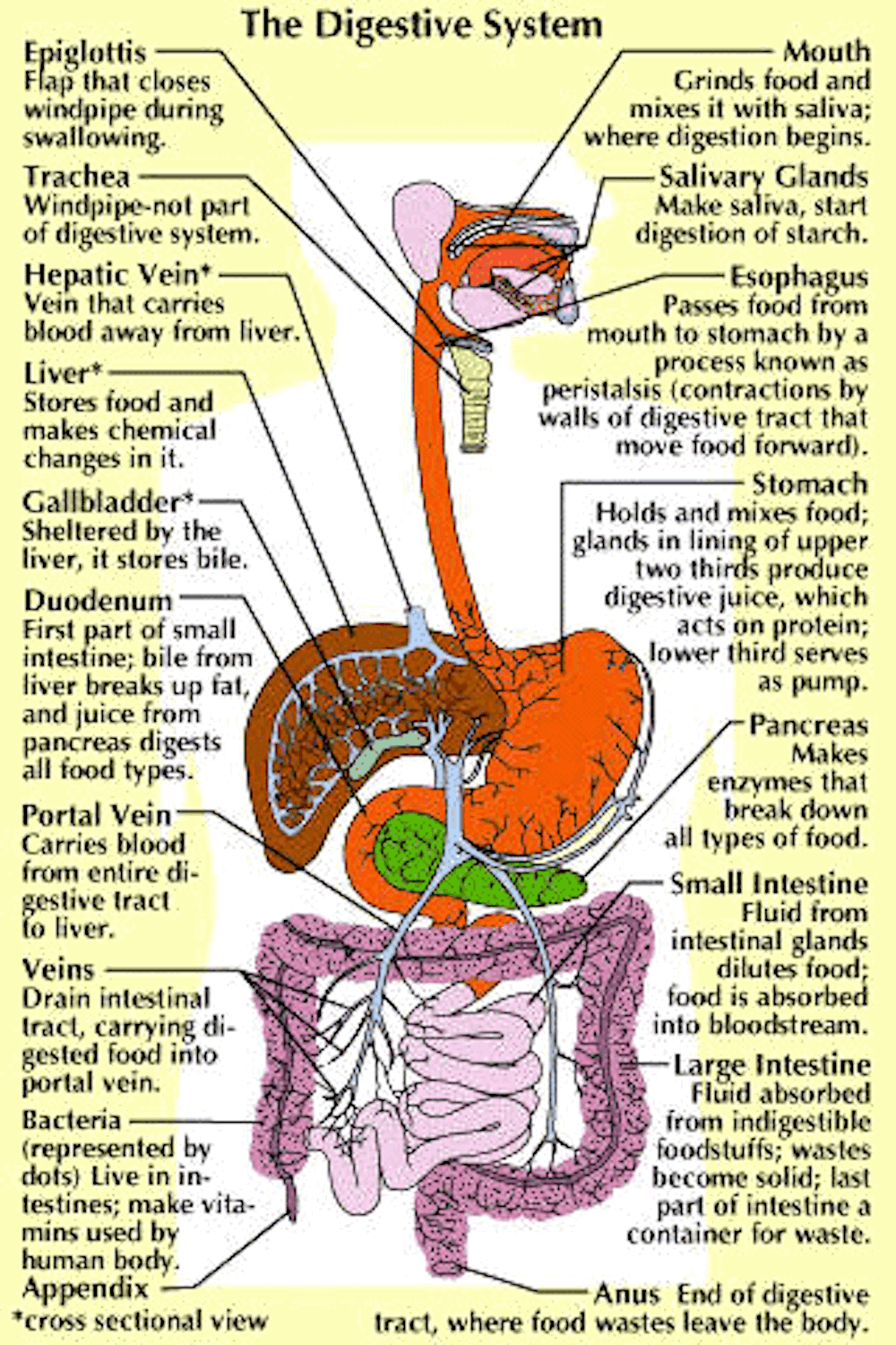
Parts and Function of Digestive System for Med School & Nursing Students - NCLEX Quiz
Venn diagram - Wikipedia A Venn diagram may also be called a set diagram or logic diagram. It is a diagram that shows all possible logical relations between a finite collection of different sets. These diagrams depict elements as points in the plane, and sets as regions inside closed curves. A Venn diagram consists of multiple overlapping closed curves, usually circles ...

Muscle Cells - Types of Cells in the Body
Labeled diagram of the human kidney royalty-free images - Shutterstock Labeled diagram of the human kidney royalty-free images 186 labeled diagram of the human kidney stock photos, vectors, and illustrations are available royalty-free. See labeled diagram of the human kidney stock video clips Image type Orientation People Artists Sort by Healthcare and Medical Anatomy Diseases, Viruses, and Disorders kidney medicine
Heart Diagram – 15+ Free Printable Word, Excel, EPS, PSD Template Download | Free & Premium ...
Cell diagram with labels - Graph Diagram Cell Diagram With Labels This human anatomy diagram with labels depicts and explains the details and or parts of the Cell Diagram With Labels. Human anatomy diagrams and charts show internal organs, body systems, cells, conditions, sickness and symptoms information and/or tips to ensure one lives in good health.
Print Exercise 7 flashcards | Easy Notecards
Cell transcriptomic atlas of the non-human primate Macaca ... Apr 13, 2022 · Studying tissue composition and function in non-human primates (NHPs) is crucial to understand the nature of our own species. Here we present a large-scale cell transcriptomic atlas that ...

25 Human Cell Diagram To Label - Wiring Database 2020
A Well-labelled Diagram Of Animal Cell With Explanation - BYJUS Well-Labelled Diagram of Animal Cell The Cell Organelles are membrane-bound, present within the cells. There are various organelles present within the cell and are classified into three categories based on the presence or absence of membrane. Listed below are the Cell Organelles of an animal cell along with their functions.
Post a Comment for "45 diagram of a human cell with labels"