38 diagram of the human heart with labels
13+ Heart Diagram Templates - Sample, Example, Format Download Color Heart Diagram Sample Format Free Download. cdhb.health.nz This colored heart diagram is a graphic representation of the organ which can be used for presentations and videos about the subject of human heart. The picture is in a coloured format and is available for a free download. Free Download. How to Draw the Internal Structure of the Heart (with Pictures) - wikiHow 1. To find a good diagram, go to Google Images, and type in "The Internal Structure of the Human Heart". Find an image that displays the entire heart, and click on it to enlarge it. 2. Find a piece of paper and something to draw with. Start with the pulmonary veins.
› male-human-anatomy-diagramMale Human Anatomy Diagram Pictures, Images and Stock Photos Pacemaker Diagram Cross section of a human heart with pacemaker fitted, showing the major arteries and veins. This is an EPS 10 vector illustration and includes a high resolution JPEG. male human anatomy diagram stock illustrations

Diagram of the human heart with labels
› heart › picture-of-the-heartHuman Heart (Anatomy): Diagram, Function, Chambers, Location ... Cardiomyopathy: A disease of heart muscle in which the heart is abnormally enlarged, thickened, and/or stiffened. As a result, the heart's ability to pump blood is weakened. As a result, the heart ... Simple Heart Diagram Labeling Activity (Teacher-Made) - Twinkl This simple heart diagram with labels activity will help your pupils begin to understand the heart, what it does and the different parts that comprise it. The resource comes with two different diagrams of the heart; one with labels attached, and one blank diagram with the labels at the bottom for students to complete themselves. Ideal as an introductory lesson on the heart and how it ... en.wikipedia.org › wiki › File:Diagram_of_the_humanFile:Diagram of the human heart (cropped).svg - Wikipedia Diagram of the human heart, created by Wapcaplet in Sodipodi. Cropped by Yaddah to remove white space (this cropping is not the same as Wapcaplet's original crop). English: Diagram of the human heart
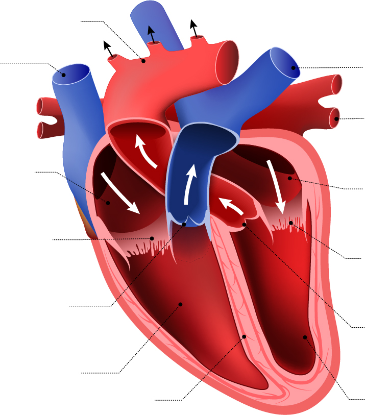
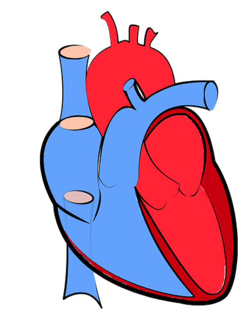
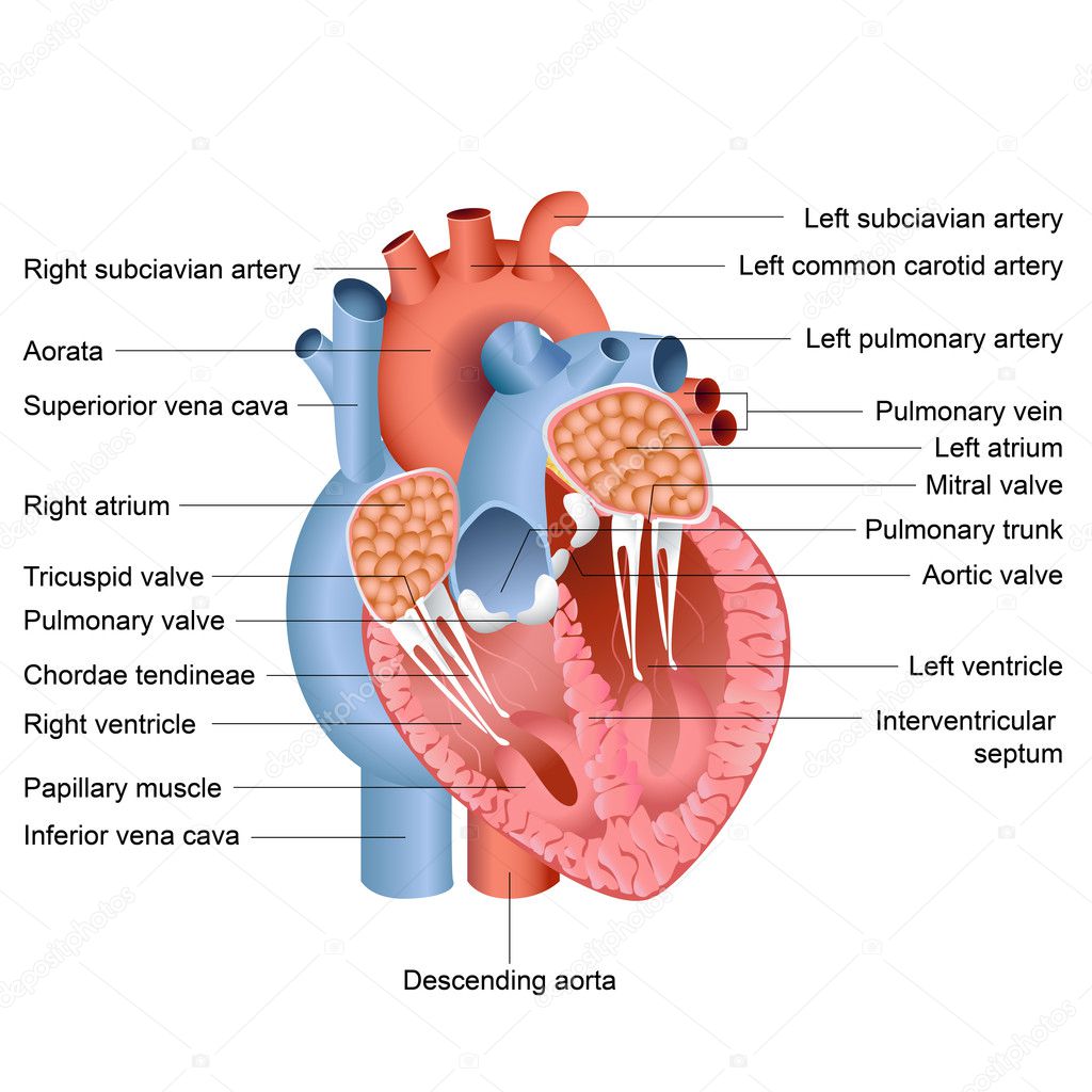
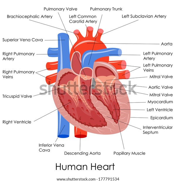
Diagram of the human heart with labels. Human Heart - Diagram and Anatomy of the Heart - Innerbody The heart functions by pumping blood both to the lungs and to the systems of the body. To prevent blood from flowing backwards or "regurgitating" back into the heart, a system of one-way valves are present in the heart. The heart valves can be broken down into two types: atrioventricular and semilunar valves. Atrioventricular valves. The atrioventricular (AV) valves are located in the middle of the heart between the atria and ventricles and only allow blood to flow from the atria into ... 667 Human Heart Diagram Premium High Res Photos Browse 667 human heart diagram stock photos and images available, or search for heart illustration or pulmonary artery to find more great stock photos and pictures. heart illustration. pulmonary artery. kidney diagram. File : Diagram of the human heart (no labels).svg - Wikimedia File:Diagram of the human heart (no labels).svg. Size of this PNG preview of this SVG file: 498 × 599 pixels. Other resolutions: 199 × 240 pixels | 399 × 480 pixels | 639 × 768 pixels | 851 × 1,024 pixels | 1,703 × 2,048 pixels | 533 × 641 pixels. KS2 The Heart Diagram QR Labelling Activity (teacher made) - Twinkl This heart diagram for children is a detailed and accurate illustration of a human heart, with labels for each section. There are also different colours to represent oxygenated (red) and deoxygenated blood (blue).
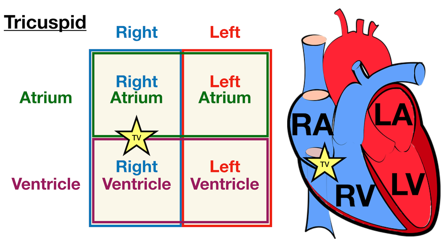
heart diagram with labels The Heart | ClipArt ETC etc.usf.edu. heart diagram labeled unlabeled human worksheet clipart simple anatomy label brain clip circulation system coronary cliparts etc pulmonary usf right. Anatomy & Physiology biologycorner.com. cat anatomy dissection arteries vessels circulatory system heart veins physiology quizlet brachiocephalic slides ... Heart Diagram for Kids - Bodytomy As you can see in the diagram of the heart, that heart is divided in four chambers, namely, right atrium, left atrium, right ventricle and left ventricle. Each of the chambers is separated by a muscle wall known as Septum. The left side of the heart receives oxygen rich blood from the lungs and pumps it out the whole body. Heart Anatomy: Labeled Diagram, Structures, Blood Flow ... - EZmed There are 4 chambers, labeled 1-4 on the diagram below. To help simplify things, we can convert the heart into a square. We will then divide that square into 4 different boxes which will represent the 4 chambers of the heart. The boxes are numbered to correlate with the labeled chambers on the cartoon diagram. View fullsize Human Heart with Labels on White Background stock photo Description Computer generated image of human heart, major arteries and veins with labeled anatomy, isolated on white background 1 credit Essentials collection for this image $4 with a 1-month subscription (10 Essentials images for $40) Continue with purchase View plans and pricing Includes our standard license. Add an extended license.
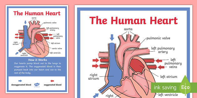
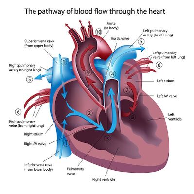
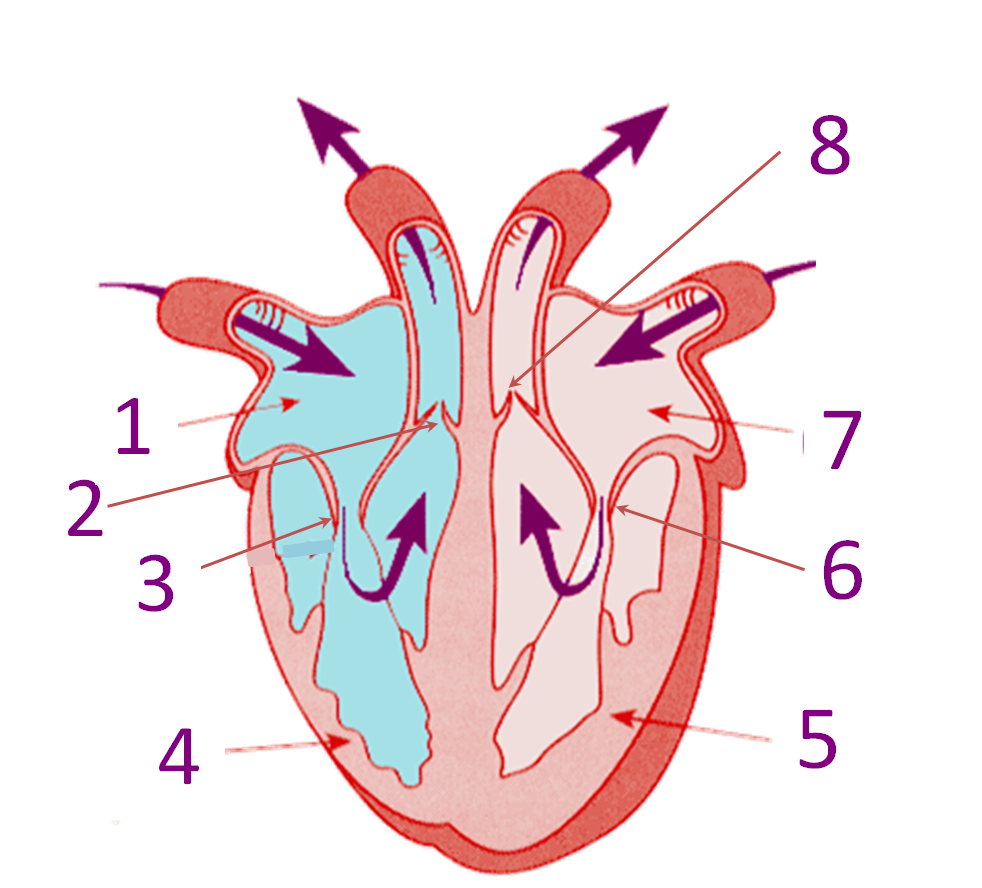
Circulatory System Diagram | New Health Advisor Heart: Human heart is a powerful pumping organ that pushes out the oxygenated blood from the left ventricle to be distributed to the periphery; whereas right heart collects deoxygenated blood ... There are different types of circulatory system diagrams; some have labels while others don't. The color blue stands for deoxygenated blood while ... Human Heart Diagram - Human Body Pictures - Science for Kids Photo description: This is an excellent human heart diagram which uses different colors to show different parts and also labels a number of important heart component such as the aorta, pulmonary artery, pulmonary vein, left atrium, right atrium, left ventricle, right ventricle, inferior vena cava and superior vena cava among others. Label the heart - Teaching resources - Wordwall Label the heart GCSE biology Labelled diagram. by 17r2hige. 10X Label the Heart diagram Labelled diagram. by Kpatel1. Label The Diagram of The Heart Labelled diagram. by Eadams4. KS2 Science Living things. Label The Diagram of The Heart Labelled diagram. by Btaylor11. Human Heart Diagram, with labelling and Function Let's understand the four chambers of the heart with a labeled Simple Heart Diagram. Human Heart Structure Diagram The heart is the main and crucial organ of the human circulatory system. The human heart is conical. The human heart structure is complex due to its function. Let,s give a look at the heart structure.
Sketch Of Human Heart Anatomy With Hand Written Labels Stock ...
› photos › diagram-of-bodyDiagram Of Body Organs Female Pics Stock Photos, Pictures ... Human internal organs Internal organs in woman and man body. Brain, stomach, heart, kidney, medical icon in female and male silhouette. Digestive, respiratory, cardiovascular systems. Anatomy poster vector illustration. diagram of body organs female pics stock illustrations

Human Heart Diagram Without Labels | Human heart diagram ...
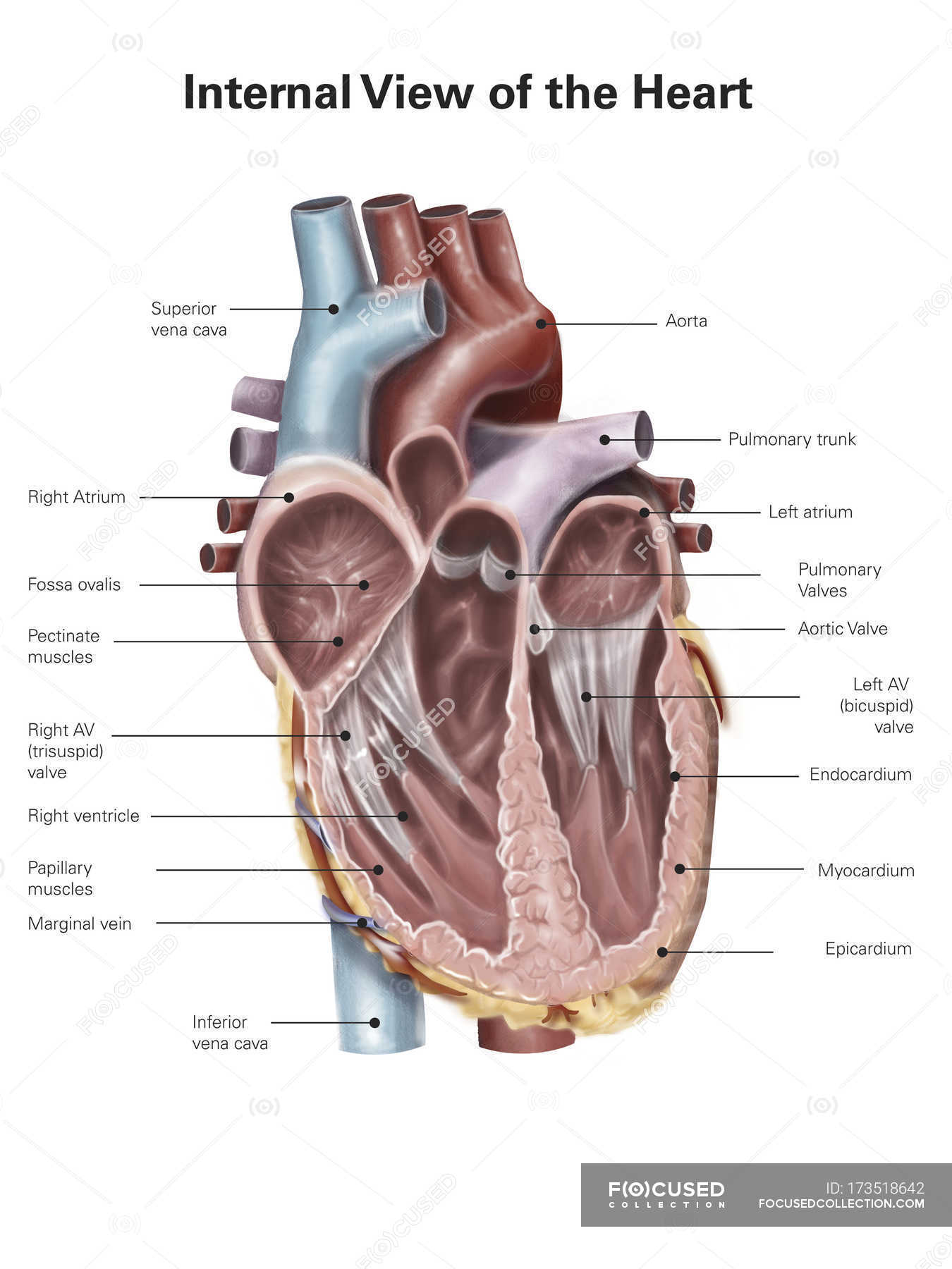
Diagram of Human Heart and Blood Circulation in It A heart diagram labeled will provide plenty of information about the structure of your heart, including the wall of your heart. The wall of the heart has three different layers, such as the Myocardium, the Epicardium, and the Endocardium. Here's more about these three layers. Epicardium
Label the Human Heart | eCampusOntario H5P Studio
Diagram of the human heart royalty-free images - Shutterstock Diagram of the human heart royalty-free images 14,996 diagram of the human heart stock photos, vectors, and illustrations are available royalty-free. See diagram of the human heart stock video clips Image type Orientation People Artists Sort by Popular Anatomy Healthcare and Medical Icons and Graphics Diseases, Viruses, and Disorders

Draw a diagram of the sectional view of human seminiferous ...
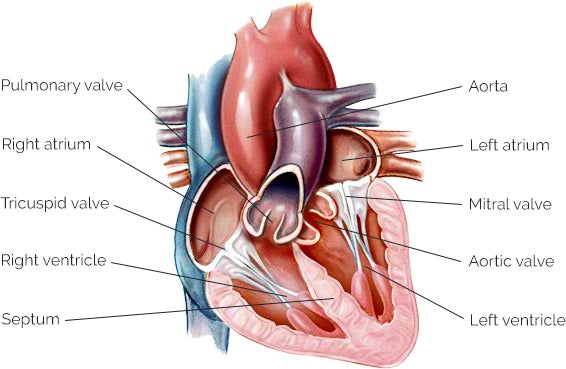
Heart Diagram with Labels and Detailed Explanation - BYJUS Well-Labelled Diagram of Heart. The heart is made up of four chambers: The upper two chambers of the heart are called auricles. The lower two chambers of the heart are called ventricles. The heart wall is made up of three layers: The outer layer of the heart wall is called epicardium. The middle layer of the heart wall is called myocardium. The inner layer of the heart wall is called endocardium.
Labelled Heart Display Poster | Primary Resources | Twinkl
en.wikipedia.org › wiki › File:Heart_diagram-enFile:Heart diagram-en.svg - Wikipedia You are free: to share – to copy, distribute and transmit the work; to remix – to adapt the work; Under the following conditions: attribution – You must give appropriate credit, provide a link to the license, and indicate if changes were made.

Cross Section Of Heart With Labels On White Background Stock ...
Human Heart Diagram Labeled | Science Trends Anatomy Of The Heart. The human heart usually weighs somewhere between 10 to 12 ounces in men and between 8 to 10 ounces in women, and in terms of size is roughly the size of the fist. The heart has four different chambers: the left and right ventricles and the left and right atriums. The chambers of the heart and the valves that regulate blood flow to them are considered the plumbing of the heart.
Label the heart — Science Learning Hub
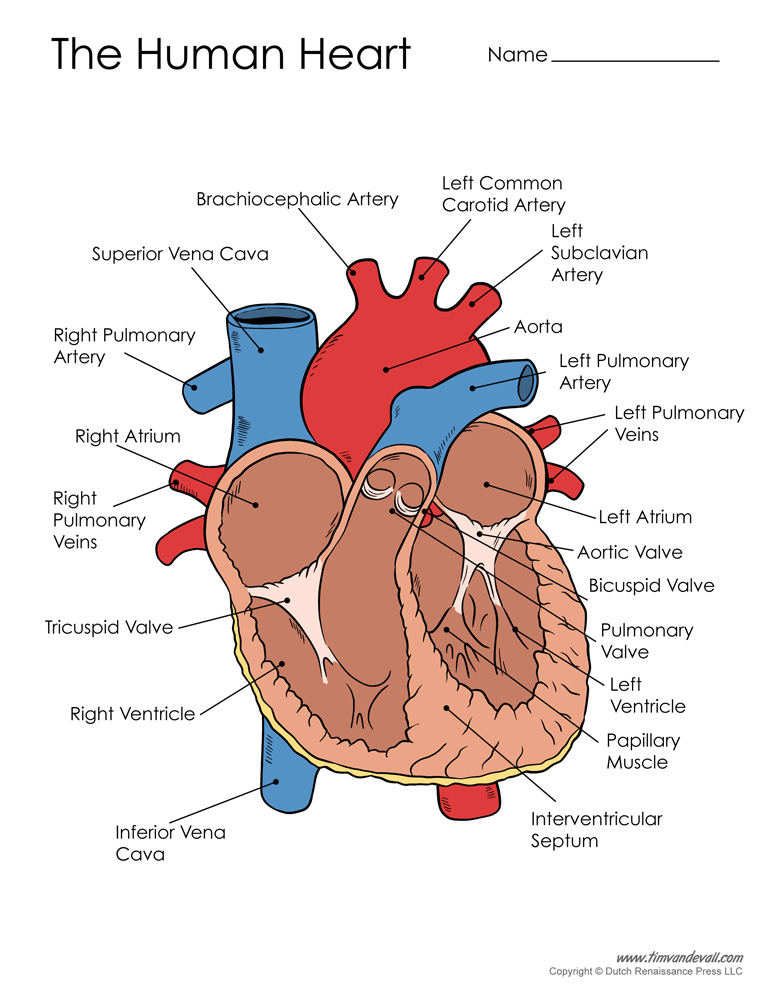
The Human Heart Labeling Worksheet (Teacher-Made) - Twinkl The human heart is a muscle made up of four chambers, these are: Two lower chambers - the left and right ventricles. It's also made up of four valves - these are known as the tricuspid, pulmonary, mitral and aortic valves. With this heart diagram without labels, you can familiarise your students with all the correct terms and help them ...
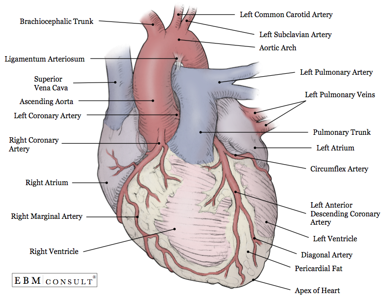
Anatomy: Heart (External)
A Labeled Diagram of the Human Heart You Really Need to See The human heart, comprises four chambers: right atrium, left atrium, right ventricle and left ventricle. The two upper chambers are called the left and the right atria, and the two lower chambers are known as the left and the right ventricles. The two atria and ventricles are separated from each other by a muscle wall called 'septum'.
Heart Anatomy: Labeled Diagram, Structures, Blood Flow ...
Heart Diagram - 15+ Free Printable Word, Excel, EPS, PSD Template ... 99+ FREE & Premium Heart Drawings - Download NOW Beautifully Designed, Easily Editable Templates to Get your Work Done Faster & Smarter. Free Download Label The Parts Of The Heart depts.washington.edu | Having the heart diagram for studies or for scientific purpose has been made easy through this template.
![Anatomy of the Heart: Structures and Blood Flow [Cardiology Made Easy]](https://i.ytimg.com/vi/1b4V09HzhBw/maxresdefault.jpg)
Anatomy of the Heart: Structures and Blood Flow [Cardiology Made Easy]
commons.wikimedia.org › wiki › File:Diagram_of_theFile : Diagram of the human heart (cropped).svg - Wikimedia Sep 29, 2022 · English: Diagram of the human heart 1. Superior vena cava 2. 4. Mitral valve 5. Aortic valve 6. Left ventricle 7. Right ventricle 8. Left atrium 9. Right atrium 10. Aorta 11. Pulmonary v

Human Heart: Label the diagram 1 worksheet
Circulatory System Diagram - Cardiovascular System and Blood ... Circulatory system diagrams are visual representations of the circulatory system, also referred to as the cardiovascular system. It is comprised of three parts: the pulmonary circulation, coronary circulation, and systemic circulation. The main function of the circulatory system is to circulate blood, which carries oxygen and nutrients ...

Human Heart Circulatory System Diagram Chart Medical Educational Science Class Anatomy Corazon Veins Arteries Labels White Wood Framed Art Poster ...
Human Heart Diagram Without Labels - Labelling Worksheet - Twinkl The human heart is a muscle made up of four chambers, these are: Two upper chambers - the left atrium and right atrium Two lower chambers - the left and right ventricles. It's also made up of four valves - these are known as the tricuspid, pulmonary, mitral and aortic valves.

Diagram of a human heart. | Download Scientific Diagram
Human Heart Diagram Pictures, Images and Stock Photos Cross Section of Heart with Labels on White Background Computer generated image of a sagittal cross section view of a human heart, showing chambers, major arteries and veins with anatomy labels. human heart diagram stock pictures, royalty-free photos & images

4,141 Human Heart Diagram Stock Photos, Pictures & Royalty ...
Spinal Cord Diagram with Detailed Illustrations and Clear Labels - BYJUS The spinal cord is one of the most important structures in the human body. It is the most important structure for any vertebrate. Anatomically, the spinal cord is made up of nervous tissue and is integrated into the spinal column of the backbone. Main Article: Spinal Cord - Anatomy, Structure, Function, and Spinal Cord Nerves; Also Read:

File:Diagram of the human heart.svg - Wikimedia Commons
› 1-label-the-heartLabel the heart — Science Learning Hub In this interactive, you can label parts of the human heart. Drag and drop the text labels onto the boxes next to the diagram. Selecting or hovering over a box will highlight each area in the diagram. pulmonary vein. semilunar valve. right ventricle. right atrium. vena cava. left atrium.

Anatomy of a Human Heart
en.wikipedia.org › wiki › File:Diagram_of_the_humanFile:Diagram of the human heart (cropped).svg - Wikipedia Diagram of the human heart, created by Wapcaplet in Sodipodi. Cropped by Yaddah to remove white space (this cropping is not the same as Wapcaplet's original crop). English: Diagram of the human heart

Colorful Hand Drawn Illustration Of Human Heart Anatomy Stock ...
Simple Heart Diagram Labeling Activity (Teacher-Made) - Twinkl This simple heart diagram with labels activity will help your pupils begin to understand the heart, what it does and the different parts that comprise it. The resource comes with two different diagrams of the heart; one with labels attached, and one blank diagram with the labels at the bottom for students to complete themselves. Ideal as an introductory lesson on the heart and how it ...
Human heart with labels — cross section, anatomy - Stock ...
› heart › picture-of-the-heartHuman Heart (Anatomy): Diagram, Function, Chambers, Location ... Cardiomyopathy: A disease of heart muscle in which the heart is abnormally enlarged, thickened, and/or stiffened. As a result, the heart's ability to pump blood is weakened. As a result, the heart ...

Human Heart Circulatory System Diagram Chart Medical Educational Science Class Anatomy Corazon Veins Arteries Labels Black Wood Framed Art Poster ...

Draw a diagram of the front view of human heart and label two ...

3d heart labeled - Google Search | Heart diagram, Human heart ...

Diagram of Human Heart Anatomy. Vector Illustration Stock ...

How to Draw the Internal Structure of the Heart (with Pictures)

Human heart with label free image download

File:Diagram of the human heart (cropped).svg - Wikimedia Commons
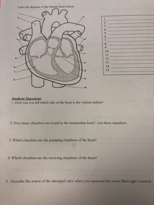
Solved Label the diagram of the human heart below. - ivono ...
Heart Anatomy: Labeled Diagram, Structures, Blood Flow ...
Anatomy of the Human Heart - Physiopedia

Human Heart Diagram Anatomy Diagram Educational Chart Thick Paper Sign Print Picture 8x12
human-heart-diagram – Tim's Printables
Heart anatomy | YourHeartValve
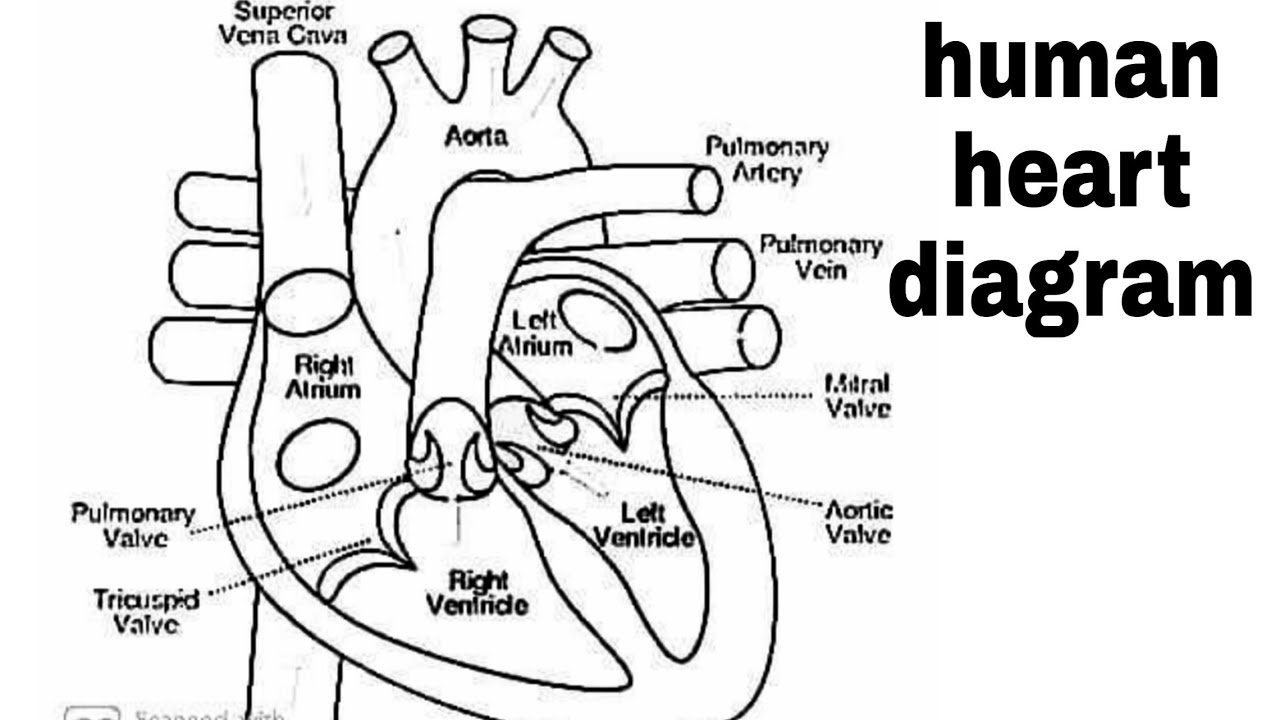
Diagram of heart/How to draw human heart easily/human heart/human heart diagram/labelled human heart
Heart Anatomy Stock Vector Image by ©stockshoppe #9978141
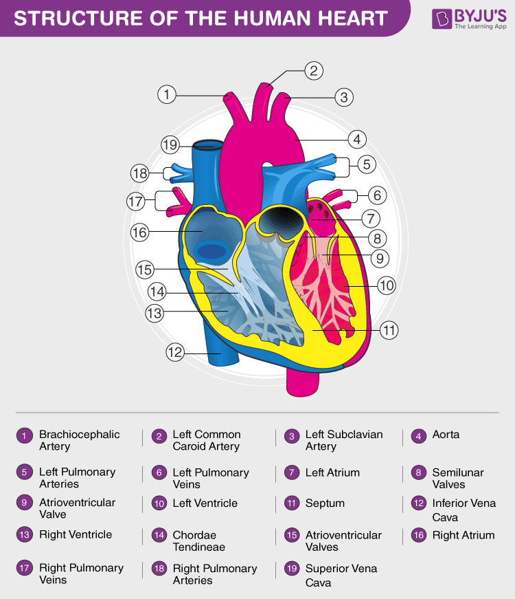
Heart Diagram with Labels and Detailed Explanation
Parts Of The Heart - ProProfs Quiz

heart | Structure, Function, Diagram, Anatomy, & Facts ...
Vector Illustration Diagram Human Heart Anatomy Stock Vector ...

parts of the heart outside - Clip Art Library
Post a Comment for "38 diagram of the human heart with labels"