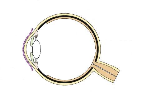
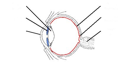
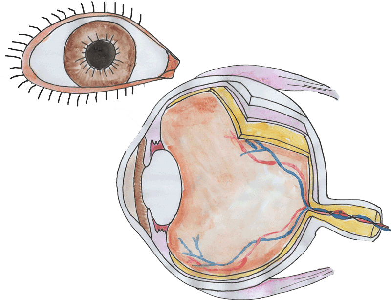
43 diagram of the human eye without labels
Eye Anatomy: Parts of the Eye and How We See Here is a tour of the eye starting from the outside, going in through the front and working to the back. Eye Anatomy: Parts of the Eye Outside the Eyeball. The eye sits in a protective bony socket called the orbit. Six extraocular muscles in the orbit are attached to the eye. These muscles move the eye up and down, side to side, and rotate the eye. File:Diagram of human eye without labels.svg - Wikimedia The following 37 pages use this file: User:Magog the Ogre/Multilingual legend/2021 June 21-30; File:Diagram of human eye without labels.svg; File:Oeil2.jpg
View your billing reports and cost trends - Google Cloud 2 päivää sitten · Labels: Labels are key/value pairs you attach to resource usage (for example, Compute Engine, Cloud Storage, or Google Kubernetes Engine).To filter usage by label, follow these steps: Expand the Labels section.; Select the label Key.; Select the Value under that key you want to filter on (the default is all values under the selected key).; To add another label with a …
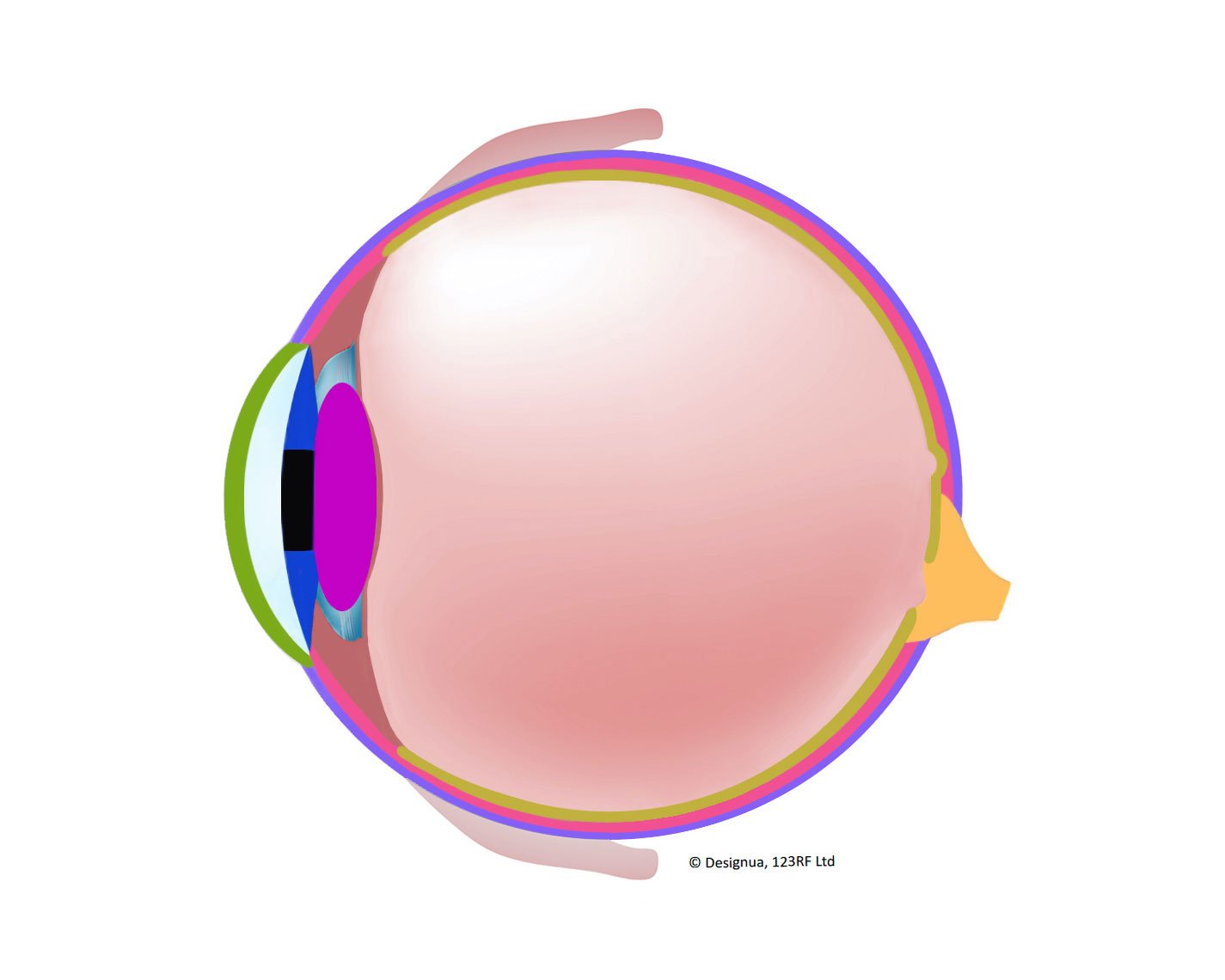
Diagram of the human eye without labels
Search PROSPERO - University of York PROSPERO accepts registrations for systematic reviews, rapid reviews and umbrella reviews. PROSPERO does not accept scoping reviews or literature scans.Sibling PROSPERO sites registers systematic reviews of human studies and systematic reviews of animal studies.. Before registering a new systematic review, check PROSPERO and the resources on COVID-END to … 6,819 Human eye diagram Images, Stock Photos & Vectors | Shutterstock Find Human eye diagram stock images in HD and millions of other royalty-free stock photos, illustrations and vectors in the Shutterstock collection. Thousands of new, high-quality pictures added every day. Human Ear Diagram - Bodytomy The Structure of Human Ear. Helix: It is the prominent outer rim of the external ear. Antihelix: It is the cartilage curve that is situated parallel to the helix. Crus of the Helix: It is the landmark of the outer ear, situated right above the pointy protrusion known as the tragus. Auditory Ossicles: The three small bones in the middle ear ...
Diagram of the human eye without labels. What Does the Eye Look Like? - Diagram of the Eye | Harvard Eye Associates Vitreous Gel: A thick, transparent liquid that fills the center of the eye. It is mostly water and gives the eye its form and shape. Our eyes are vital for seeing the world around us. Keep them healthy by maintaining regular vision exams. Contact Harvard Eye Associates at 949-951-2020 or harvardeye.com to schedule an appointment today. label the ear worksheet Picture Front Of The Eye Without Labels Clipart 20 Free Cliparts clipground.com. eye human diagram worksheet eyeball learning layers without anatomy labels parts eyes worksheets clipart science structure grade clipground body structures. 14 Best Images Of Ear Hearing Worksheets - Listening Ear Craft Template Structure and Functions of Human Eye with labelled Diagram - BYJUS The human eye is a roughly spherical organ, responsible for perceiving visual stimuli. It is enclosed within the eye sockets in the skull and is anchored down by muscles within the sockets. Anatomically, the eye comprises two components fused into one; hence, it does not possess a perfect spherical shape. Call Girls in Delhi & Escort Service in Delhi - Aditi Ghosh I am Aditi Ghosh a call girl in Delhi I am a prostitute who serves you for your enjoyment, I provide both incall and outcall in hotel room services and I do not display my profession to the general public, I am usually work in an organization which I love brothels though and have been working independently as a escort in Delhi for the past 2 years.
Aerocity Escorts & Escort Service in Aerocity @ vvipescort.com Aerocity Escorts @9831443300 provides the best Escort Service in Aerocity. If you are looking for VIP Independnet Escorts in Aerocity and Call Girls at best price then call us.. label the eye worksheet 15 Best Images Of Anatomy On Human Eye Worksheet - Human Eye Diagram frog diagram dissection worksheet anatomy human muscle heart pelvic system labeled eye pectoral worksheeto lessons chapter unlabeled via lab circulatory Basic Eye Anatomy Quiz eye anatomy diagram basic quiz game purposegames Anatomy of the eye: Quizzes and diagrams | Kenhub Here you can see all of the main structures in this area. Spend some time reviewing the name and location of each one, then try to label the eye yourself - without peeking! - using the eye diagram (blank) below. Unlabeled diagram of the eye Click below to download our free unlabeled diagram of the eye. human eye diagram with labels Human Skeleton Blank Clip Art at Clker.com - vector clip art online. 8 Pics about Human Skeleton Blank Clip Art at Clker.com - vector clip art online : Human Eye Diagrams with the Unlabeled, picture front of the eye without labels clipart - Clipground and also Male Reproductive System | Free Images at Clker.com - vector clip art.
human eye diagram with labels Eye With Labels Clip Art at Clker.com - vector clip art online, royalty. 10 Images about Eye With Labels Clip Art at Clker.com - vector clip art online, royalty : Muscles of the Human Eyeball | ClipArt ETC, 35 Label The Structure Of The Eye - Labels Database 2020 and also 35 Label The Structure Of The Eye - Labels Database 2020. label the ear worksheet Picture Front Of The Eye Without Labels Clipart - Clipground clipground.com eye human diagram worksheet layers eyeball learning labels anatomy without clipart eyes parts worksheets science structure clipground grade body structures 12 Best Images Of Anatomy Human Ear Diagram Worksheet - Blank Ear Data and information visualization - Wikipedia Data and information visualization (data viz or info viz) is an interdisciplinary field that deals with the graphic representation of data and information.It is a particularly efficient way of communicating when the data or information is numerous as for example a time series.. It is also the study of visual representations of abstract data to reinforce human cognition. Principles of Epidemiology: Lesson 4, Section 4|Self-Study ... The size of the pie is related to the size of the population in that age group. Because the population in one age group is larger, 1 pie is larger than the other. This makes it difficult to compare causes of death for the 2 groups without reading the labels. Return to text. Figure 4.28b
Labelling the eye — Science Learning Hub
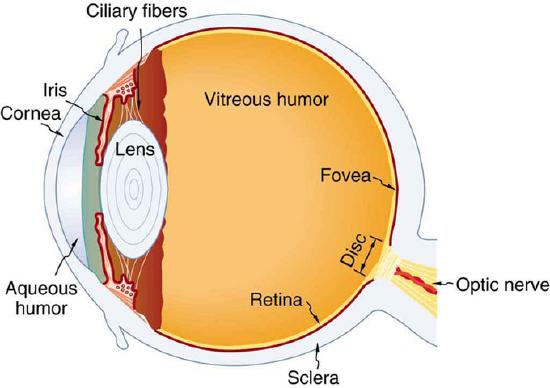
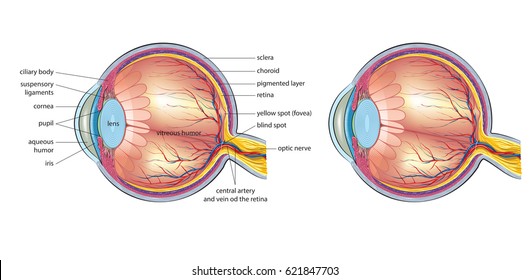
PDF Parts of the Eye - National Institutes of Health Eye Diagram Handout Author: National Eye Health Education Program of the National Eye Institute, National Institutes of Health Subject: Handout illustrating parts of the eye Keywords: parts of the eye, eye diagram, vitreous gel, iris, cornea, pupil, lens, optic nerve, macula, retina Created Date: 12/16/2011 12:39:09 PM
Anatomy of the Human Eye: Identify, Label and Color
human anatomy diagram without labels Abdominal Anatomy Male - Human Body Diagram Without Labels . abdominal netclipart. Brain Cross-section - Wait But Why waitbutwhy.com. brain section sagittal cross without anatomi human half cerebellum mid cerebrum right midbrain brainstem corpus eye mind functions structure callosum. Liam Roberts BAGD YR2: Arm Anatomy Reference

Module 1: Labeled Diagram of the Eye | Diagram of the eye ...
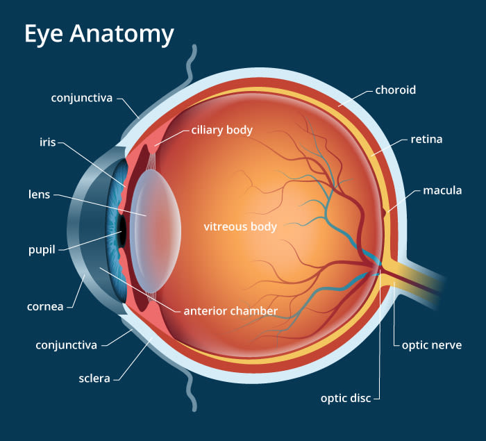
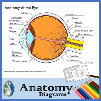
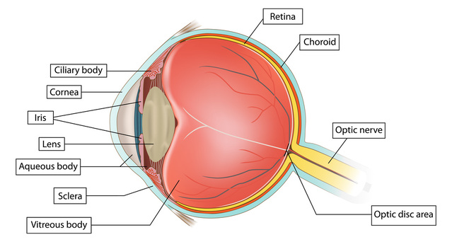
PDF Eye Anatomy Handout - National Institutes of Health of light entering the eye. Lens: The lens is a clear part of the eye behind the iris that helps to focus light, or an image, on the retina. Macula: The macula is the small, sensitive area of the retina that gives central vision. It is located in the center of the retina. Optic nerve: The optic nerve is the largest sensory nerve of the eye.

Vector Illustration Of Diagram Of Eye Anatomy With Label ...
File : Schematic diagram of the human eye en.svg - Wikimedia Diagram of the human eye in English. It shows the lower part of the right eye after a central and horizontal section. ... Full redraw: Group labels in accordance with the "Foundational Model Explorer." Added "Macula" and "Uvea" and removed "Zonular fibres." ... File:Diagram of human eye without labels.svg; File:Figure of diplopia perception ...

Diagram of the Eye Side View No Labels | Diagram of the eye ...
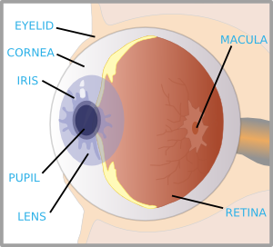
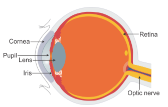
The Eyes (Human Anatomy): Diagram, Optic Nerve, Iris, Cornea ... - WebMD Your eye is a slightly asymmetrical globe, about an inch in diameter. The front part (what you see in the mirror) includes: Iris: the colored part; Cornea: a clear dome over the iris; Pupil: the ...
Label Eye Printout - EnchantedLearning.com
IIS 7.5 详细错误 - 404.0 - Not Found | Human eye diagram, Eye anatomy ... Human Eye Anatomy Worksheet coloring page from Anatomy category. Select from 64000 printable crafts of cartoons, nature, animals, Bible and many more. A collection of embroidery and cross stitch patterns from independent designers. Sublime Stitching, Subversive Cross Stitch, Studio MME, and others.
13,197 Eye Diagram Images, Stock Photos & Vectors | Shutterstock
Labelled Diagram of Human Eye, Explanation and Function - VEDANTU The human eye is a part of the sensory nervous system. Labeled Diagram of Human Eye The eyes of all mammals consist of a non-image-forming photosensitive ganglion within the retina which receives light, adjusts the dimensions of the pupil, regulates the availability of melatonin hormones, and also entertains the body clock.

Draw a Diagram of the Human Eye as Seen in a Vertical Section ...
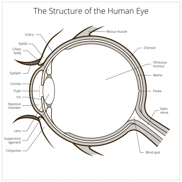
Eye Diagram With Labels and detailed description - BYJUS A brief description of the eye along with a well-labelled diagram is given below for reference. Well-Labelled Diagram of Eye The anterior chamber of the eye is the space between the cornea and the iris and is filled with a lubricating fluid, aqueous humour. The vascular layer of the eye, known as the choroid contains the connective tissue.
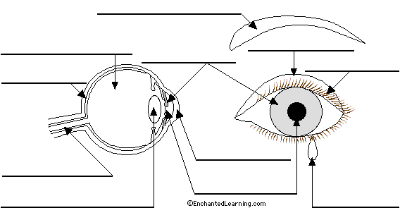
Label Eye Printout #2 - EnchantedLearning.com
Making Content Usable for People with Cognitive and Learning Disabilities This document is for people who make web content (web pages) and web applications. It gives advice on how to make content usable for people with cognitive and learning disabilities. This includes, but is not limited to: cognitive disabilities, learning disabilities (LD), neurodiversity, intellectual disabilities, and specific learning disabilities.

Free art print of Diagram of human eye anatomy with label
human face diagram and label human face diagram and label picture front of the eye without labels clipart - Clipground. 9 Images about picture front of the eye without labels clipart - Clipground : muscle neck diagram blank labels - Google Search | Neck muscle anatomy, New Page 1 [classroom.sdmesa.edu] and also picture front of the eye without labels clipart - Clipground.

Cross Section of a Human Eye Diagram Black and White ...
Eye diagram by Firkin | Human eye diagram, Diagram of the eye, Eye ... A great way to show your students how the eye works! Give your students an interactive way to learn about the structure of the human eye. Students make their eye model using the colourful parts working alone or in small groups. They then use their model to look at the structure of the eye, how it works and learn how visual defects are corrected.
Eye Anatomy: A Closer Look At the Parts of the Eye
Human eye - Wikipedia The human eye is a sensory organ, part of the sensory nervous system, that reacts to visible light and allows humans to use visual information for various purposes including seeing things, keeping balance, and maintaining circadian rhythm.. The eye can be considered as a living optical device.It is approximately spherical in shape, with its outer layers, such as the outermost, white …

3d Image Render Of Diagram Of Eye Anatomy With Label For ...
Human Heart (Anatomy): Diagram, Function, Chambers, Location in ... - WebMD WebMD's Heart Anatomy Page provides a detailed image of the heart and provides information on heart conditions, tests, and treatments.

File:Diagram of human eye without labels.svg - Wikimedia Commons
Technicolor Home Page Vantiva . Vantiva, formerly known as Technicolor, is composed of businesses with leading positions in their respective markets and with solid growth fundamentals: Vantiva Broadband, Video, IoT and Supply Chain Solutions.
Eye With Labels Clip Art at Clker.com - vector clip art ...
respiratory tract diagram without labels respiratory system label smoking ppt powerpoint presentation. 32 Label The Respiratory System Quiz - Labels 2021 documentdowu.blogspot.com. quiz. Respiratory system diagram to label. Respiratory system diagram grade circulatory systems body human label sciences natural science labels breathing main siyavula za components gr9 mstworkbooks.

Diagram human eye anatomy with label Royalty Free Vector
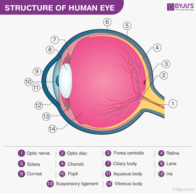
The Human Eye - Diagram, Parts, Working, Function and Work of The Lens The cornea, iris, pupil, and lens make up the front of the eye, which focuses the image onto the retina. The light-sensitive membrane that covers the back of the eye is known as the retina. This membrane is made up of millions of nerve cells that clump together behind the eye to form the optic nerve, a huge nerve. The Human Eye
Human Eye - Definition, Structure, Function, Parts, Diagram
Label Parts of the Human Eye - University of Dayton Parts of the Eye. Select the correct label for each part of the eye. The image is taken from above the left eye. Click on the Score button to see how you did. Incorrect answers will be marked in red. ...
FREE! - The Human Eye Labeling Activity
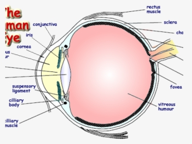
Eye Anatomy: 16 Parts of the Eye & Their Functions - Vision Center The following are parts of the human eyes and their functions: 1. Conjunctiva. The conjunctiva is the membrane covering the sclera (white portion of your eye). The conjunctiva also covers the interior of your eyelids. Conjunctivitis, often known as pink eye, occurs when this thin membrane becomes inflamed or swollen.
Eye Diagram With Labels - ClipArt Best
Human Body Diagram - Bodytomy ☛ The human eye has the ability to differentiate between 400+ shades of gray, and what's more, it can identify approximately 10 million colors. ☛ Your ears never sleep. Sound is received even while you are asleep; it's the brain that does not process them.

Anatomy of the Human Eye
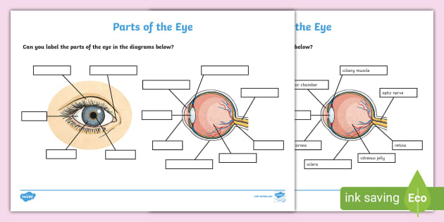
FREE! - The Human Eye Labelling Activity - Twinkl In this resource, you’ll find a 2-page PDF that is easy to download, print out, and use immediately with your class. The first page is a labelling exercise with two diagrams of the human eye. One is a view from the outside, and the other is a more detailed cross-section. Challenge learners to label the parts of the eye diagram. On the second page, you’ll find a set of answers showing ...
Anatomy of the eye: Quizzes and diagrams | Kenhub
Eye Anatomy: A Closer Look At the Parts of the Eye - All About Vision In a number of ways, the human eye works much like a digital camera: Light is focused primarily by the cornea — the clear front surface of the eye, which acts like a camera lens. The iris of the eye functions like the diaphragm of a camera, controlling the amount of light reaching the back of the eye by automatically adjusting the size of the ...
25.1: The Human Eye - Physics LibreTexts
PHSchool.com Retirement–Prentice Hall–Savvas Learning … PHSchool.com was retired due to Adobe’s decision to stop supporting Flash in 2020. Please contact Savvas Learning Company for product support.
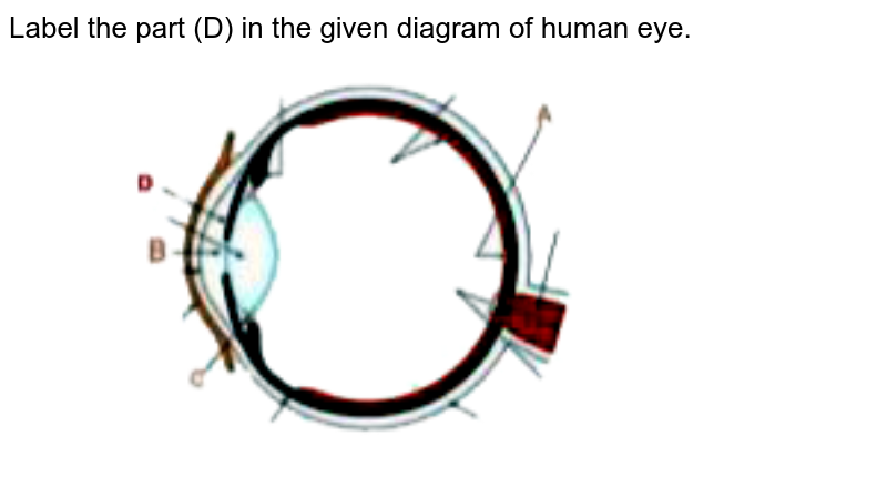
Label the part (D) in the given diagram of human eye.
Human Ear Diagram - Bodytomy The Structure of Human Ear. Helix: It is the prominent outer rim of the external ear. Antihelix: It is the cartilage curve that is situated parallel to the helix. Crus of the Helix: It is the landmark of the outer ear, situated right above the pointy protrusion known as the tragus. Auditory Ossicles: The three small bones in the middle ear ...

Human Eye Diagram Stock Illustration - Download Image Now ...
6,819 Human eye diagram Images, Stock Photos & Vectors | Shutterstock Find Human eye diagram stock images in HD and millions of other royalty-free stock photos, illustrations and vectors in the Shutterstock collection. Thousands of new, high-quality pictures added every day.

Diagram of human eye anatomy with label illustration. | CanStock
Search PROSPERO - University of York PROSPERO accepts registrations for systematic reviews, rapid reviews and umbrella reviews. PROSPERO does not accept scoping reviews or literature scans.Sibling PROSPERO sites registers systematic reviews of human studies and systematic reviews of animal studies.. Before registering a new systematic review, check PROSPERO and the resources on COVID-END to …
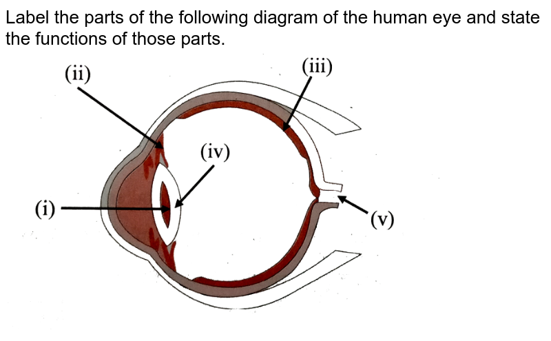
Label the parts of the following diagram of the human eye and ...
13,197 Eye Diagram Images, Stock Photos & Vectors | Shutterstock

Draw a neat and label diagram of human eye and explain ...
Eye structure Vector Art Stock Images | Depositphotos
OLCreate: HEAT_NCD_ET_1.0 Non-Communicable Diseases ...

Rudyard.org | Eye anatomy diagram, Eye anatomy, Diagram of ...
Labelled Diagram Of Human Eye , Png Download - Label A Human ...

eye_labeling_drag_and_drop - St. Catherine of Genoa ~ St ...
Draw the structure of human eye and label any three parts of ...
Anatomy of the Eye Diagrams for Coloring/Labeling, with ...
The Internal Structure - The Human Eye

Human Biology- Chapter 15 Flashcards | Quizlet
unlabeled human eye diagram - Clip Art Library

How to draw Human Eye Diagram | #HUMAN #EYE #DIAGRAM #CBSE #NCERT class 10 #Shorts
Eyes - Layers of Learning

Structure Of Human Eye Without Label Transparent PNG ...
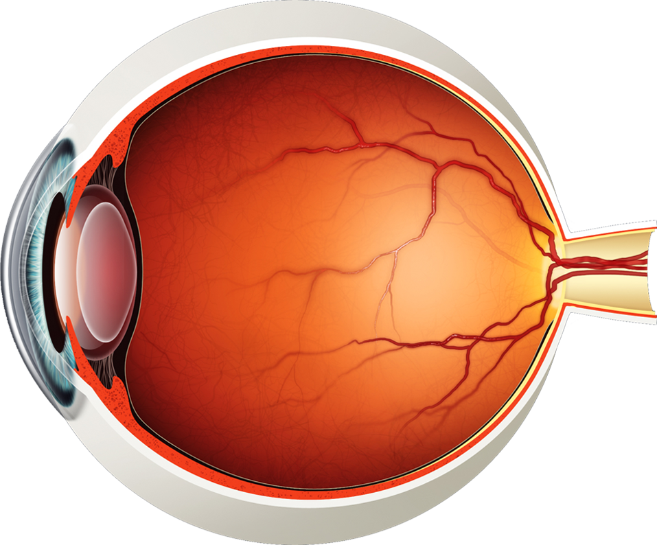
Diagram of the Eye - Lions Eye Institute

1,383 Human Eye Diagram Stock Photos, Pictures & Royalty-Free ...
Post a Comment for "43 diagram of the human eye without labels"