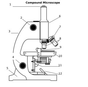
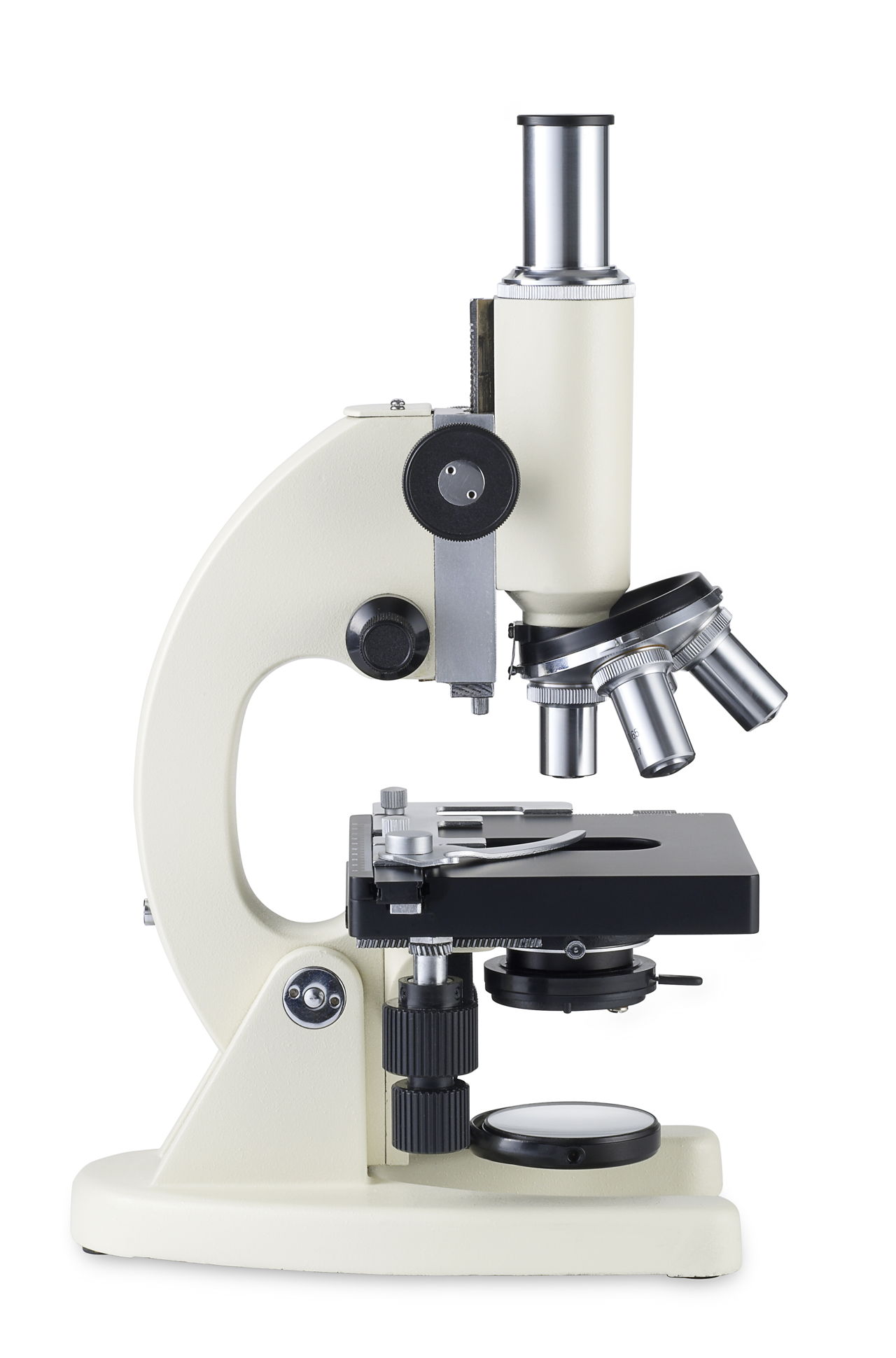
38 compound microscope diagram without labels
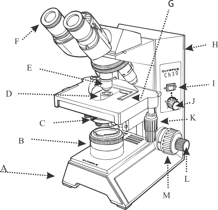
Compound Light Microscope: Everything You Need to Know A compound light microscope is a type of light microscope that uses a compound lens system, meaning, it operates through two sets of lenses to magnify the image of a specimen. It's an upright microscope that produces a two-dimensional image and has a higher magnification than a stereoscopic microscope. It also goes by a couple of other names ... Compound Microscope Parts, Functions, and Labeled Diagram Compound Microscope Definitions for Labels. Eyepiece (ocular lens) with or without Pointer: The part that is looked through at the top of the compound microscope. Eyepieces typically have a magnification between 5x & 30x. Monocular or Binocular Head: Structural support that holds & connects the eyepieces to the objective lenses.
Microscope World | Shop Microscopes For Every Application Labeling the Parts of the Microscope This activity has been designed for use in homes and schools. Each microscope layout (both blank and the version with answers) are available as PDF downloads. You can view a more in-depth review of each part of the microscope here. Download the Label the Parts of the Microscope PDF printable version here.
Compound microscope diagram without labels
Compound Microscope: Definition, Diagram, Parts, Uses, Working ... - BYJUS A compound microscope is defined as A microscope with a high resolution and uses two sets of lenses providing a 2-dimensional image of the sample. The term compound refers to the usage of more than one lens in the microscope. Also, the compound microscope is one of the types of optical microscopes. Compound Microscope- Definition, Labeled Diagram, Principle, Parts, Uses Alternatively, the magnification of the compound microscope is given by: m = D/ fo * L/fe where, D = Least distance of distinct vision (25 cm) L = Length of the microscope tube fo = Focal length of the objective lens fe = Focal length of the eye-piece lens Parts of a Compound Microscope Eyepiece And Body Tube. PDF Parts of a Microscope Printables - Homeschool Creations typical student microscope -other microscopes will vary) •Which part of the microscope rotates so another person can look through the eyepiece without needing to move the microscope ? the head •What is the magnification level on the eyepiece of a microscope?10x (see objective lens magnification to see how these work together)
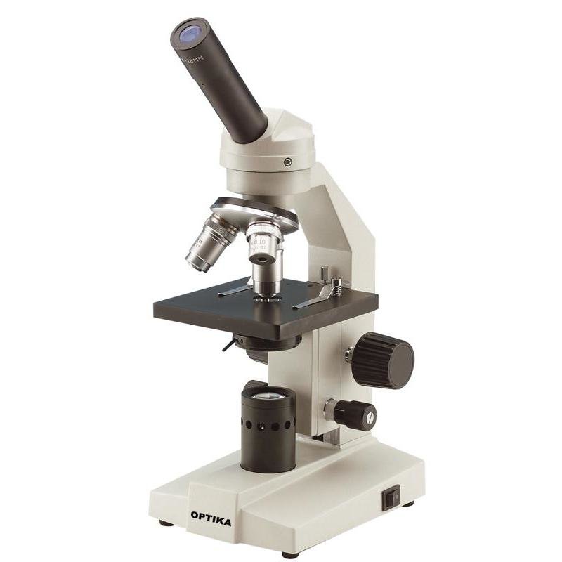
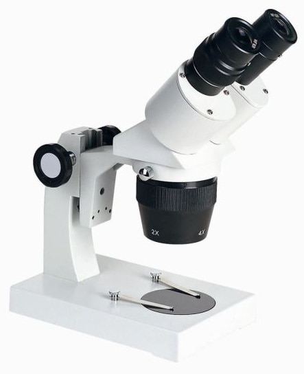
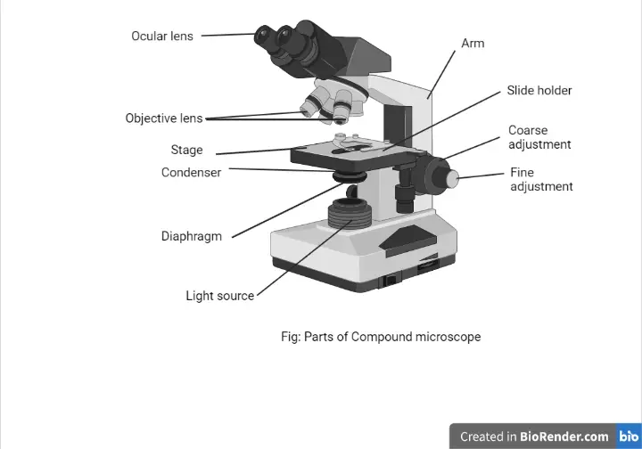
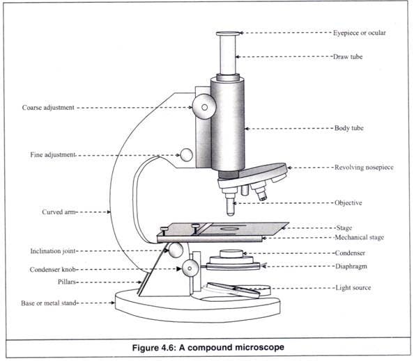
Compound microscope diagram without labels. Working Principle and Parts of a Compound Microscope (with Diagrams) It holds the stage, body tube, fine adjustment and coarse adjustment. 5. Body Tube: It is usually a vertical tube holding the eyepiece at the top and the revolving nosepiece with the objectives at the bottom. The length of the draw tube is called 'mechanical tube length' and is usually 140-180 mm (mostly 160 mm). 6. Parts of Stereo Microscope (Dissecting microscope) – labeled diagram ... The difference between Compound and Stereo (Dissecting) Microscope. Unlike a compound microscope that can only see a very thin specimen, stereo microscopes can be used for viewing almost anything you can fit under them. However, stereo microscopes offer lower magnification, typically 5x-50x, comparing to compound microscopes. PDF Basic Observation Procedures for Compound Microscopes 3. Rotate the 100X objective into position without getting the 40X objective in the oil. 4. While observing from one side of the stage, slowly, raise the stage until you see the meniscus of the oil on the specimen come in contact with the tip of the 100X objective. Now go to the eyepieces and observe as you finish focusing with the fine focus knob. 16 Parts of a Compound Microscope: Diagrams and Video Once you have an understanding of the parts of the microscope it will be much easier to navigate around and begin observing your specimen, which is the fun part! The 16 core parts of a compound microscope are: Head (Body) Arm Base Eyepiece Eyepiece tube Objective lenses Revolving Nosepiece (Turret) Rack stop Coarse adjustment knobs
Lower Secondary Science LEARNER’S BOOK 8 - Issuu 22-02-2021 · Read Lower Secondary Science LEARNER’S BOOK 8 by Cambridge University Press Education on Issuu and browse thousands of other publications on our p... K To 12 Science Grade 7 Learners Material - Module Table 2. Compounds and their constituent elements written in the food labels Food Product Compound Constituent Element Cereal Drink Chocolate candy Soy sauce Note: Please add rows as necessary 3. The elements iron and zinc are listed in the Nutrition Facts for the cereal drink. Find out from the Ingredients the source of these elements. 4. On-chip beam rotators, adiabatic mode converters, and … 07-07-2022 · Low-loss-integrated optical waveguides have indispensable roles in a wide range of significant modern technologies, such as integrated photonic chips 1,2,3, quantum applications 4,5,6,7,8,9, and ... Diagram of a Compound Microscope - Biology Discussion A bright-field or compound microscope is primarily used to enlarge or magnify the image of the object that is being viewed, which can not otherwise be seen by the naked eye. Magnification may be defined as the degree of enlargement of the image of an object provided by the microscope.
Microscope Labeling Activity | Sticky Note Labels - Classful This clear and simple microscope labeling activity is exactly what your students need to learn the parts of a compound microscope! There are five versions of microscope labeling diagrams included: ⭐Sticky Note microscope to label ⭐Microscope to label with a word bank ⭐Microscope to label without a word bank ⭐Plain microscope to label without lines ⭐Version with and without name/date ... PDF AN INTRODUCTION TO THE COMPOUND MICROSCOPE - Rowan University microscope in an upright position using both hands. **When carrying the microscope, place one hand on the base and the other hand around the arm. **DO NOT PLACE THE MICROSCOPE IN AN UPSIDE DOWN POSITION. PIECES WILL FALL OUT. **Keep microscope away from the edge of the bench, particularly when not in use. **Make sure power cords are out of the way. Looking at the Structure of Cells in the Microscope A typical animal cell is 10–20 μm in diameter, which is about one-fifth the size of the smallest particle visible to the naked eye. It was not until good light microscopes became available in the early part of the nineteenth century that all plant and animal tissues were discovered to be aggregates of individual cells. This discovery, proposed as the cell doctrine by Schleiden and … Compound Microscope: Parts of Compound Microscope - BYJUS The parts of the compound microscope can be categorized into: Mechanical parts; Optical parts (A) Mechanical Parts of a Compound Microscope. 1. Foot or base. It is a U-shaped structure and supports the entire weight of the compound microscope. 2. Pillar. It is a vertical projection. This stands by resting on the base and supports the stage. 3. Arm
Label a microscope - Teaching resources
Microscope Parts and Functions The compound microscope is more complicated than just a microscope with more than one lens. Read on. ... It also allows the specimen to be labeled, transported, and stored without damage. Stage: The flat platform where the slide is placed. Stage clips: ... Basics of a Compound Microscope. Diagram/Parts/Functions of a Compound Microscope.
Parts of a Microscope Quiz
Electron microscope - Wikipedia An electron microscope is a microscope that uses a beam of accelerated electrons as a source of illumination. As the wavelength of an electron can be up to 100,000 times shorter than that of visible light photons, electron microscopes have a higher resolving power than light microscopes and can reveal the structure of smaller objects.. Electron microscopes use shaped magnetic …

The Microscope
Parts of a Compound Microscope and Their Functions - NotesHippo Compound microscope magnification is determined by multiplying the eyepiece and objective powers. When viewed through a 5X eyepiece with a 10X objective, an item is magnified 5 x 10=50 times. The magnification is 10 x 45 = 450 times when using a 10X eyepiece and a 45X objective. How to Use the Compound Microscope

Microscope Labeling
Compound Microscope - Types, Parts, Diagram, Functions and Uses A compound microscope captures an inverted image of the specimen because every time the light passes through the lens, the image's direction is flipped. The image always ends up inverted from the original. So, if you move the sample to the left, it moves in the right direction. Image 18: A comparison image between a simple and compound microscope.
Simple Microscope - Parts, Functions, Diagram and Labelling ...
ALEX | Alabama Learning Exchange Subject: Digital Literacy and Computer Science (4), Science (4) Title: Using Code to Create an Animated Animal Description: Students will use the free online coding program, Scratch, to learn the basics of coding and how to use blocks and animations to create an animated animal. Students will show how an animated animal will receive, process, and respond to information …

Light Microscope- Definition, Principle, Types, Parts ...
(PDF) general-chemistry.pdf | Sumit Banerjee - Academia.edu general-chemistry.pdf

Biology - labeling a compound microscope Diagram | Quizlet
A Study of the Microscope and its Functions With a Labeled Diagram ... Compound Microscope Diagram The compound microscope uses light for illumination. Some compound microscopes make use of natural light, whereas others have an illuminator attached to the base. The specimen is placed on the stage and observed through different lenses of the microscope, which have varying magnification powers.

microscope | Types, Parts, History, Diagram, & Facts | Britannica
Labelled Diagram of Compound Microscope The below mentioned article provides a labelled diagram of compound microscope. Part # 1. The Stand: The stand is made up of a heavy foot which carries a curved inclinable limb or arm bearing the body tube. The foot is generally horse shoe-shaped structure (Fig. 2) which rests on table top or any other surface on which the microscope in kept.

Label the microscope — Science Learning Hub
Parts of the Microscope with Labeling (also Free Printouts) Parts of the Microscope with Labeling (also Free Printouts) By Editorial Team March 7, 2022 A microscope is one of the invaluable tools in the laboratory setting. It is used to observe things that cannot be seen by the naked eye. Table of Contents 1. Eyepiece 2. Body tube/Head 3. Turret/Nose piece 4. Objective lenses 5. Knobs (fine and coarse) 6.
Parts of a Microscope with Their Functions – Microbe Online
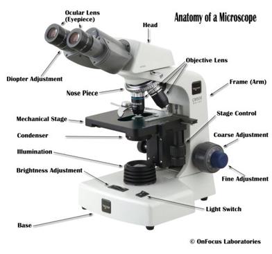
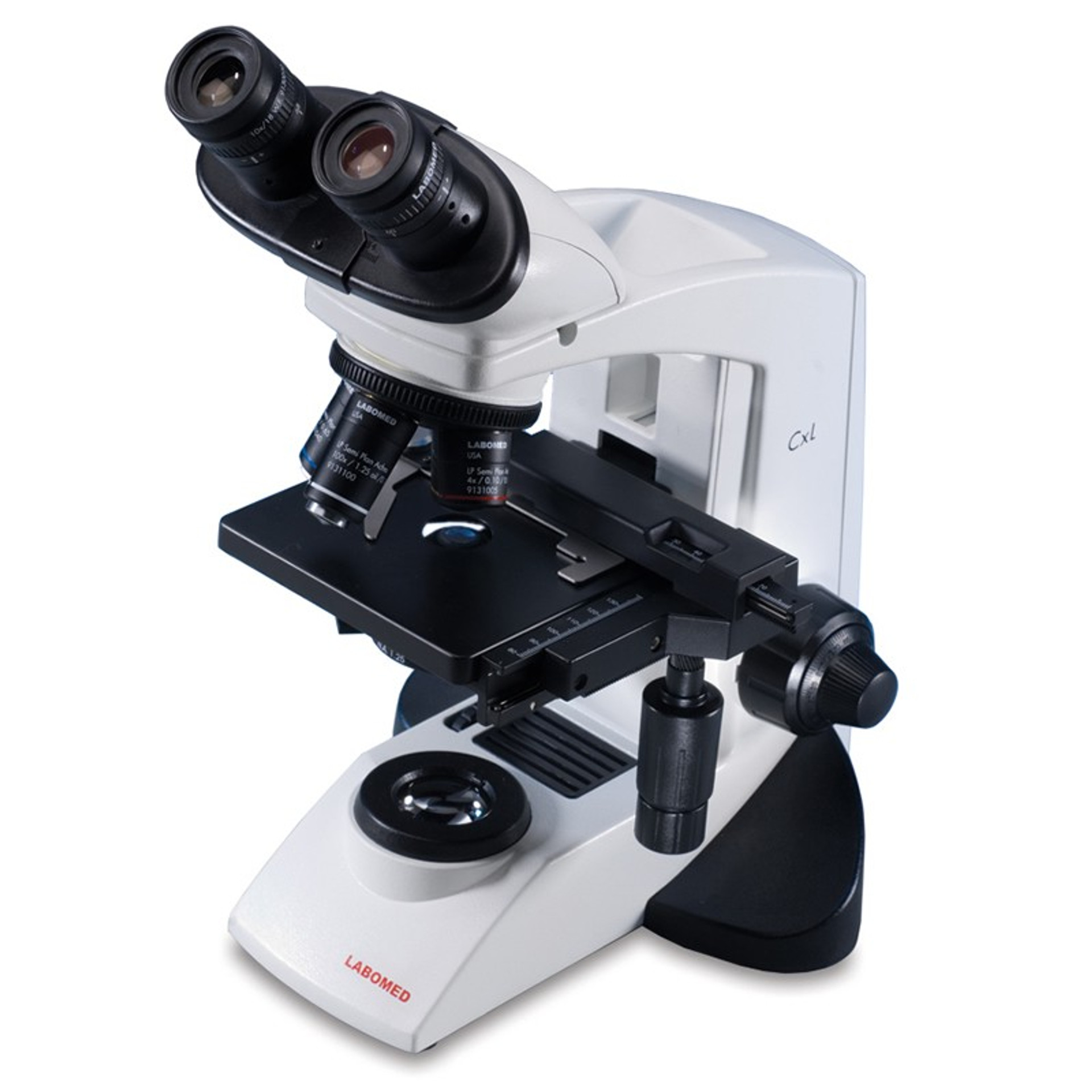
Binocular Microscope Anatomy - Parts and Functions with a Labeled Diagram Now, I will discuss the details anatomy of the light compound microscope with the labeled diagram. Why it is called binocular: because it has two ocular lenses or an eyepiece on the head that attaches to the objective lens, this ocular lens magnifies the image produced by the objective lens. Binocular microscope parts and functions
16 Parts of a Compound Microscope: Diagrams and Video ...
Inorganic Chemistry 4th edition, Catherine Housecroft Purple acid phosphatases (PAPs) are a group of metallohydrolases that contain a dinuclear Fe(III)M(II) center (M(II) = Fe, Mn, Zn) in the active site and are able to catalyze the hydrolysis of a variety of phosphoric acid esters.

Compound Light Microscope Labeling Diagram | Quizlet
Solved Label the image of a compound light microscope using - Chegg Expert Answer. 100% (17 ratings) Transcribed image text: Label the image of a compound light microscope using the terms provided.

Compound Microscope Parts – Labeled Diagram and their ...
Label the microscope — Science Learning Hub In this interactive, you can label the different parts of a microscope. Use this with the Microscope parts activity to help students identify and label the main parts of a microscope and then describe their functions. Drag and drop the text labels onto the microscope diagram.

Compound Microscope- Definition, Labeled Diagram, Principle ...
Label A Microscope Teaching Resources | Teachers Pay Teachers This is a set of 3 tiered readings. Students will read a passage about the how to use a compound light microscope. Students will use textual evidence to answer questions and label the different parts of the microscope. It also allows students to gain prior knowledge about the compound microscope. Version A provides the most support for students.

Microscope Diagram Labeled, Unlabeled and Blank | Parts of a ...
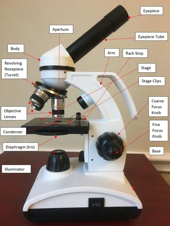
Microscope, Microscope Parts, Labeled Diagram, and Functions The Iris Diaphragm is located above the condenser lens and below the microscope stage. The different sized holes in the diaphragm helps to vary the size of the cone and intensity of light that is projected upward into the slide. However, there is no set rule regarding which setting to use for a particular power.
Microscope Labeling
Compound Microscope Parts - Labeled Diagram and their Functions The term "compound" refers to the microscope having more than one lens. Basically, compound microscopes generate magnified images through an aligned pair of the objective lens and the ocular lens. In contrast, "simple microscopes" have only one convex lens and function more like glass magnifiers.

Microscope Diagram Labeled, Unlabeled and Blank | Parts of a ...
Parts of a Compound Microscope (And their Functions) - Scope Detective List of Microscope Parts and their Functions. 1. Ocular Tubes (Monocular, Binocular & Trinocular) The ocular tubes, are to tubes that lead from the head of the microscope out to your eyes. On the end of the ocular tubes are usually interchangeable eyepieces (commonly 10X and 20X) that increase magnification.
Introduction to the Microscope Lab Activity
Fluorescence - Wikipedia Fluorescence is the emission of light by a substance that has absorbed light or other electromagnetic radiation.It is a form of luminescence.In most cases, the emitted light has a longer wavelength, and therefore a lower photon energy, than the absorbed radiation.A perceptible example of fluorescence occurs when the absorbed radiation is in the ultraviolet …

Compound Microscope Parts – Labeled Diagram and their ...
Microscope Labeling Game - PurposeGames.com About this Quiz. This is an online quiz called Microscope Labeling Game. There is a printable worksheet available for download here so you can take the quiz with pen and paper. This quiz has tags. Click on the tags below to find other quizzes on the same subject. Science.
Compound Microscope Review - ppt download
Parts of a microscope with functions and labeled diagram - Microbe Notes Figure: Diagram of parts of a microscope There are three structural parts of the microscope i.e. head, base, and arm. Head - This is also known as the body. It carries the optical parts in the upper part of the microscope. Base - It acts as microscopes support. It also carries microscopic illuminators.
Microscope
Compound Microscope Parts Made Easy - Microscope Detective Not labeled in the above diagram but you can see it as a lightly glowing circle in the middle of the slide. (Above the reflection of the objective lens, which shows as a black circle). Illuminator - The illuminator provides the light for the microscope. It's usually a light bulb that's located underneath the stage.
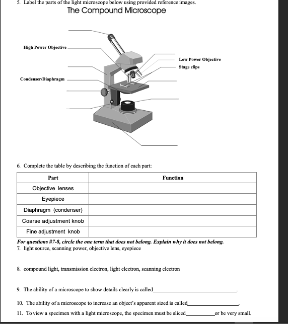
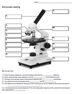
Solved 5. Label the parts of the light microscope below ...
PDF Parts of a Microscope Printables - Homeschool Creations typical student microscope -other microscopes will vary) •Which part of the microscope rotates so another person can look through the eyepiece without needing to move the microscope ? the head •What is the magnification level on the eyepiece of a microscope?10x (see objective lens magnification to see how these work together)
compound-microscope-unlabeled.jpg
Compound Microscope- Definition, Labeled Diagram, Principle, Parts, Uses Alternatively, the magnification of the compound microscope is given by: m = D/ fo * L/fe where, D = Least distance of distinct vision (25 cm) L = Length of the microscope tube fo = Focal length of the objective lens fe = Focal length of the eye-piece lens Parts of a Compound Microscope Eyepiece And Body Tube.
Building a Fully-Functional Microscope From Lego Bricks and a ...
Compound Microscope: Definition, Diagram, Parts, Uses, Working ... - BYJUS A compound microscope is defined as A microscope with a high resolution and uses two sets of lenses providing a 2-dimensional image of the sample. The term compound refers to the usage of more than one lens in the microscope. Also, the compound microscope is one of the types of optical microscopes.

Compound Microscope Parts
Microscope Parts and Functions
Color the Microscope Parts

Diagram of a Compound Microscope

Compound Microscope Parts – Labeled Diagram and their ...
Compound Microscope Clip Art at Clker.com - vector clip art ...
Understanding the Compound Microscope Parts and its Functions ...
Compound Microscope Parts, Functions, and Labeled Diagram ...

Compound Microscope – Diagram (Parts labelled), Principle and ...

Carl Zeiss Microscopy Optical microscope Worksheet Diagram ...
Free Microscope Drawing, Download Free Microscope Drawing png ...

Microscope labeled diagram

Compound Microscope Parts – Labeled Diagram and their ...
The Compound Light Microscope Label the following parts on ...
Post a Comment for "38 compound microscope diagram without labels"